Range of non-invasive angiology services
Overview and information for referring physicians
Duplex
The ultrasound devices we use represent the state of the art in ultrasound technology and offer numerous advantages for vascular medicine. With their outstanding image quality, advanced image processing functions and automated measurement tools, they provide valuable support for precise diagnostics and the efficient performance of vascular examinations.
Duplex of the carotids
Duplex sonography of the carotids is an important preventive measure for the early detection of carotid stenosis, which can significantly increase the risk of strokes. It is also a suitable, gentle and time-saving diagnostic method for detecting any atherosclerotic vascular processes before, for example, initiating lipid-lowering therapy. Early detection and treatment of carotid stenosis can help to reduce the risk of stroke and improve the patient prognosis.
Indications for screening
According to the guidelines of the German Society for Ultrasound in Medicine (DEGUM), the European Stroke Organisation (ESO), the European Society of Cardiology (ESC) and the American Heart Association (AHA), screening for carotid stenosis is recommended for
- People with symptomatic cerebrovascular disease
- Asymptomatic high-risk individuals such as patients with risk factors such as hypertension, hyperlipidaemia, diabetes mellitus, smoking or a positive family history of atherosclerotic disease.
- Screening can be considered prior to cardiovascular surgery in order to reduce the perioperative risk.
Performance and diagnostic criteria
- B-scan sonography: visualisation of the vessel wall and identification of plaques.
- Colour Doppler sonography: Visualisation of the blood flow and detection of stenoses using colour flow imaging.
- Spectral Doppler sonography: Measurement of the blood flow velocity to quantify the degree of stenosis, primarily using the NASCET classification
Sensitivity and specificity
Duplex sonography has a high diagnostic accuracy, which has been demonstrated in many studies.
Sensitivity: 85-90% for the detection of high-grade carotid stenosis (≥70% stenosis).
Specificity: 90-95% for differentiating between significant and non-significant stenosis.
Preparation of the patients
For optimal image quality and accurate diagnosis, please advise patients of the following preparation measures:
- Clothing: The patient should wear loose, comfortable clothing and avoid jewellery or tight-fitting collars.
- Medication: The patient should take their prescribed medication as usual, unless otherwise instructed.
Duplex of the aorta
Duplex of the aorta and abdominal vessels: Duplex sonography of the abdominal aorta and abdominal vessels is an essential diagnostic method for the assessment of vascular pathologies in the abdominal area. This examination is particularly important for screening the abdominal aorta for a possible aneurysm, such as suspected Dunbar's syndrome (median arcuate ligament syndrome, MALS). Dunbar's syndrome is caused by compression of the celiac trunk by the median arcuate ligament of the diaphragm and leads to abdominal pain, especially after eating.
Indications for screening
According to the current guidelines of the German Society for Ultrasound in Medicine (DEGUM), the German Society for Vascular Surgery and Vascular Medicine (DGG) and international specialist societies such as the European Society for Vascular Surgery (ESVS) and the US Preventive Services Task Force (USPSTF), screening for abdominal aortic aneurysms is recommended for
- Men 65 years of age and older: Men are at higher risk for developing a BAA, and the risk increases with age.
- Women aged 65 and over with risk factors: Especially if they are smokers or have a family history of aneurysms.
- People with a family history: Patients with a positive family history of BAA or other aneurysms.
- Smokers or former smokers: As smoking is a significant risk factor for the development of a BAA.
Indications for testing for Dunbar syndrome/MALS
Duplex ultrasonography of the abdominal aorta and abdominal vessels is indicated in patients with:
- Chronic or postprandial abdominal pain
- Suspected abdominal vascular stenoses or aneurysms
- Weight loss of unknown origin
- Nausea or vomiting after meals
- Continued suspicion of Dunbar's syndrome after exclusion of other gastrointestinal causes (inconspicuous gastrointestinal endoscopy)
Measurements and diagnostic criteria
Duplex sonography enables non-invasive assessment of the abdominal aorta and abdominal vessels with regard to
- Diameter and morphology
- blood flow velocity
- flow profile with/without provocation manoeuvre
- post-stenotic turbulence
Patient preparation
In order to optimise sonifiability and accurate diagnostics, we ask you to inform patients of the following preparation measures:
- Fasting: The patient does not necessarily have to be fasting. However, care should be taken to ensure that no flatulent foods (pulses, cabbage, onions, etc.) are consumed on the day of the examination or the day before. If possible, the patient should also only drink clear water on the day of the examination and avoid caffeinated or carbonated drinks.
- Medication can be taken as usual
Advantages of the examination
Duplex sonography of the abdominal aorta and abdominal vessels offers several decisive advantages:
- No radiation exposure or X-ray contrast media required, which makes the examination safe and, above all, the diagnostic method of choice for screening and follow-up checks
- Possibility of direct assessment of the vessels and blood flow in real time.
- High sensitivity and specificity: Precise detection of stenoses, aneurysms and other pathological changes.
- Cost-effectiveness: Compared to other imaging procedures such as CT angiography, duplex sonography is more cost-effective and still diagnostically valuable.
The early and precise diagnosis of abdominal vascular pathologies, especially Dunbar syndrome, can lead to more effective treatment planning and improved patient prognosis. Please do not hesitate to contact us if you have any questions or require detailed information on making an appointment.
FKDS of the veins / suspected DVT / CVI
FKDS of the veins for the diagnosis of suspected thrombosis (DVT) or chronic venous insufficiency (CVI):
Colour-coded duplex sonography (FKDS) is the preferred imaging method for diagnosing venous thrombosis, especially deep vein thrombosis (DVT). This method enables a non-invasive, accurate and rapid assessment of the venous vessels and provides both anatomical and haemodynamic information.
We use a high-frequency linear ultrasound probe, with which compression is also performed in layers from proximal to distal. We apply the following diagnostic criteria in order to achieve the highest possible sensitivity (>93%) and specificity (97%)
- B-scan sonography: direct visualisation of the vein wall and lumen. A thrombosis is visualised as endoluminal material within the vessel in 2 planes.
- Compression sonography: The vein is compressed by applying light pressure with the probe. A normal vein collapses completely, whereas a thrombosed vein cannot be compressed.
- Colour Doppler sonography: Assessment of the blood flow within the vein. A lack of or reduced blood flow indicates a thrombosis.
- Spectral Doppler sonography: Measurement of the flow velocity and pattern. Abnormal flow patterns (e.g. reduced or no flow) support the diagnosis.
Renal artery duplex
Renal artery duplex ultrasonography is performed to diagnose renal artery stenosis (NAST). This examination is particularly indicated for patients with
- Difficult to control or resistant hypertension, diatolic accentuated arterial hypertension or absence of a nocturnal depression in the LZ blood pressure profile
- Suspicion of renovascular hypertension
- Deterioration of kidney function without recognisable cause
- Differential diagnosis for unclear abdominal complaints
Patient preparation
In order to ensure optimum sound quality during the examination, we ask you to inform patients of the following preparation measures:
- On the day of the examination, the patient may only drink clear water. Other drinks and solid food should be avoided so as not to impair the view of the renal arteries.
- Light meals: The day before the examination, the patient should eat light meals and avoid legumes and large amounts of food to minimise gas build-up in the intestines.
Diagnostic criteria for renal artery stenosis (NAST)
During renal artery duplex ultrasonography, we use the following diagnostic criteria to assess renal artery stenosis:
- Increased blood flow velocity and a renal-aortic quotient >3.5
- Poststenotic turbulence
- Delayed early diastolic rise (acceleration time, AT): An AT of more than 100 milliseconds may indicate a significant stenosis.
- Lateral difference
Please inform patients about these preparatory measures and the importance of the examination for accurate diagnosis and treatment planning.
FKDS in case of Raynaud's syndrome
FKDS in cases of Raynaud'ssyndrome and arterial compression syndromes: In addition to capillary microscopy and photoplethysmography, we also offer a duplex sonographic examination primarily to exclude significant macroangiopathies in order to make a precise diagnosis and differentiate between primary and secondary Raynaud's syndrome. As a provocation manoeuvre supported by duplex sonography, we carry out the fist-closure test for patients with Raynaud's or examine our patients in certain positions or under other provocation manoeuvres (e.g. Adson test, Roos test for thoracic outlet syndrome, diagnosis of entrapment syndromes) during the ultrasound.
Symptoms
Patients often complain of flank pain, haematuria (blood in the urine), proteinuria, varicocele (in men) and occasionally abdominal pain together with ‘misdiagnosed’ PCO syndrome.
Clinical examination
The physical examination can provide evidence of a varicocele, particularly on the left side in men.
Imaging procedures
Doppler ultrasound: If the blood flow assessment reveals compression of the left renal vein together with evidence of increased blood flow velocity in the compressed renal vein and possible reflux into the gonadal veins, an imaging scan is planned to confirm the diagnosis and plan treatment (CT angiography (CTA) or MR angiography (MRA) depending on the patient's characteristics.
Description
Cross-sectional imaging procedure that provides detailed images of the vessels and shows the anatomical relationship between the left renal vein, the aorta and the superior mesenteric artery.
Findings
Confirmation of compression of the left renal vein between the aorta and the superior mesenteric artery, dilated precompressive renal vein and collaterals.
FKDS with May-Thurner syndrome
The FKDS is a valuable tool for the initial diagnosis of May-Thurner syndrome. The visualisation of the anatomical relationship between the left common iliac vein and the right common iliac artery is essential, followed by quantification of blood flow velocity and direction to identify potential flow disturbances. Occasionally, the pre- and post-stenotic pressure gradient can be estimated non-invasively and in some cases thromboses in the affected vein can already be recognised on ultrasound.
If the findings are positive, we initiate further diagnostic measures (sectional imaging for treatment planning depending on the patient characteristics).
Photoplethysmography
- Procedure: The change in blood flow in the fingers is measured using infrared light. This helps to document vasospastic reactions.
- Speciality: This examination is often carried out in combination with thermal provocation tests.
- Duration: Appointments are made individually, usually as part of a second appointment due to the longer preparation and follow-up time
In order to achieve optimum results, we ask you to advise patients of the following preparatory measures
- No exposure to cold: The patient should take care not to expose themselves to cold for a prolonged period 24 hours before the examination.
- Avoidance of stimulants: Products containing caffeine and nicotine should be avoided at least 12 hours before the examination.
- Relaxation: The patient should relax sufficiently before the examination and avoid stress in order not to falsify the results.
Capillary microscopy
Procedure: The microcirculation in the capillaries of the nail bed is examined using a microscope. This method is particularly helpful in identifying morphological changes that indicate secondary Raynaud's syndrome.
- No exposure to cold: A few hours before the examination, the patient should ensure that the hands/feet are no longer exposed to cold.
- Please do not appear with artificial fingernails
- Avoidance of stimulants: Products containing caffeine and nicotine should be avoided at least 6 hours before the examination.
- Relaxation: The patient should relax sufficiently before the examination and avoid stress so as not to distort the results.
TcpO2 testing, especially for chronic limb ischaemia
TcpO2 measurement is a valuable tool in vascular medicine that enables precise and objective assessment of peripheral blood flow. The method is particularly helpful for patients with chronic wounds and PAD, as it allows an accurate assessment of tissue oxygenation and the chances of healing.
Compared to other diagnostic procedures, TcpO2 measurement is non-invasive, easy to perform and offers fast and objectifiable results that are scientifically established. We perform this diagnostic procedure primarily for the following questions:
- Differentiation of the severity of the circulatory disorder
- Assessment of the chances of healing chronic wounds, ulcers and diabetic foot syndromes.
- Assessment of blood flow prior to vascular surgery or amputations.
- Monitoring the effectiveness of revascularisation measures such as angioplasty or bypass operations.
Standardisierte Gehstreckenermittlung/Knöchel-Arm Index
Die standardisierte Gehstreckenmessung ist ein wichtiges diagnostisches Verfahren zur Beurteilung der peripheren arteriellen Verschlusskrankheit (pAVK). Sie ermöglicht die objektive Quantifizierung der Gehfähigkeit und die Einteilung der Krankheitsstadien, was für die Planung der Therapie und die Überwachung des Krankheitsverlaufs von großer Bedeutung ist. Die Gehstreckenmessung dient zur Ermittlung der maximalen Gehstrecke, die ein Patient ohne Pause zurücklegen kann, und der schmerzfreien Gehstrecke, bis das Auftreten von Claudicatio-Symptomen wie Schmerzen, Krämpfen oder Müdigkeit in den Beinen. Diese Daten helfen bei der Beurteilung der Schwere der pAVK und der Lebensqualität des Patienten.
Die standardisierte Messung erfolgt meist auf einem Laufband unter kontrollierten Bedingungen. Das Laufband wird auf eine konstante Geschwindigkeit eingestellt, in der Regel 3-4 km/h, mit einer leichten Steigung (ca. 10-12%). Der Patient beginnt zu gehen, und die Zeit bis zum Auftreten der ersten Symptome (schmerzfreie Gehstrecke) und bis zum Erreichen der maximal tolerierbaren Gehstrecke wird gemessen.
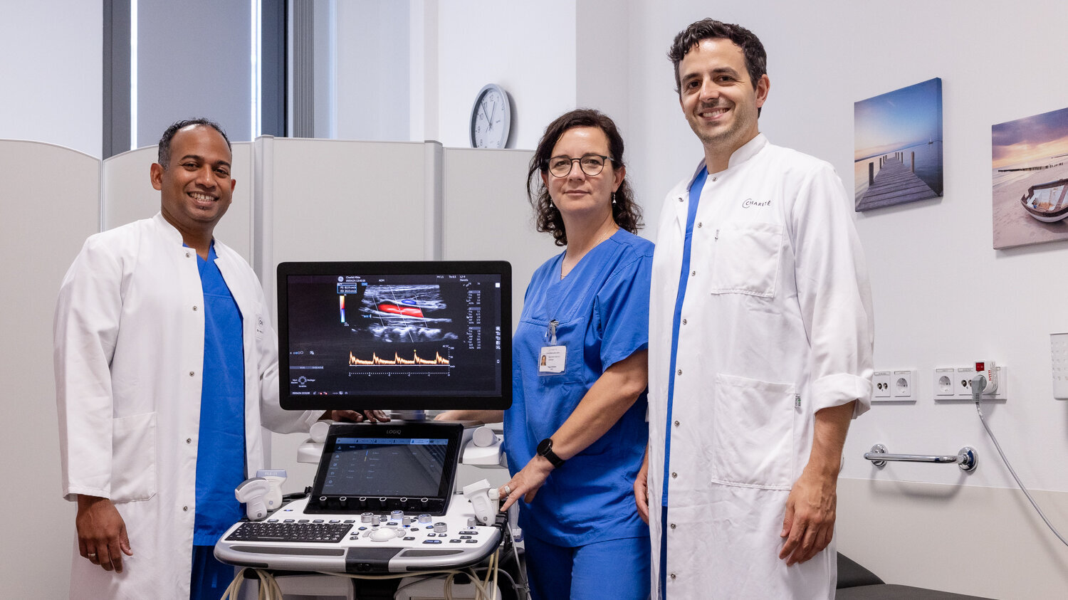
The team of the angiological function laboratory at the Department of Cardiology, Angiology and Intensive Care Medicine at the Charité Mitte campus.
The team of the angiological function laboratory at the Department of Cardiology, Angiology and Intensive Care Medicine at the Charité Mitte campus.