Management
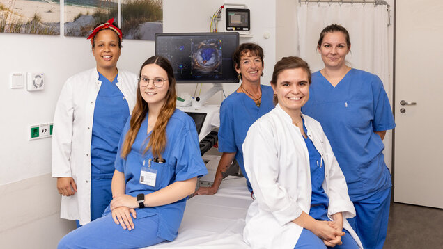
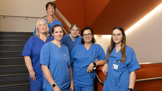
Our Department of Functional Diagnostics is headed by Elena Romero Dorta.

Management
Our Department of Functional Diagnostics is headed by Elena Romero Dorta.
Cardiological functional diagnostics (heart diagnostics)
Cardiological functional diagnostics includes various methods for examining the cardiovascular system. Echocardiography, spiroergometry, ECG, stress ECG and long-term ECG can be used to recognise diseases and prepare for important interventions and/or operations.
The focus of cardiological functional diagnostics is on echocardiography (cardiac ultrasound) in our certified echocardiography laboratory as well as various types of ECG diagnostics.
Echocardiography is one of the most important diagnostic methods in modern cardiology. The heart is examined using ultrasound waves (as with an echo sounder). Both the shape and function of the heart can be assessed.
There are various examination methods, such as transthoracic echocardiography, in which the transducer is positioned on the chest. Transoesophageal echocardiography is performed using an endoscope with a transducer at the tip. The endoscope is inserted through the oesophagus and allows the examiner to fully scan the atrial areas.
Echocardiography with its various examination techniques (B-mode, M-mode echocardiography; Doppler echocardiography; contrast echocardiography) enables the function of the heart to be diagnosed, e.g. with regard to its pumping capacity, heart size, wall movement and changes in the heart valves. Due to the accuracy of the examination results, echocardiography is an excellent examination method without radiation exposure, in which all essential structures can be visualised.
Stress echocardiography is used to search for circulatory disorders in the heart.
The examination is used to search for areas with poor blood flow following a pathological ergometry result. The stress echocardiography is performed with medication that the patient receives via an infusion into the vein. Alternatively, similar to ergometry (stress ECG), the stress can be induced on a recumbent bicycle. Like Doppler echocardiography, the examination itself is performed with the transducer on the chest.
Under stress, disturbances in the wall movement of the heart muscle in particular become visible, which indicate a lack of blood flow in this area.
The electrocardiogram (ECG) records the electrical processes that occur during cardiac activity. The recording is carried out on a millimetre paper running at a defined speed. This makes it possible to calculate the heart rate and the duration of excitation of the individual sections of the heart from the width of the individual teeth and sections. It also provides information about the position of the heart in the chest, the heart rhythm and the heart rate. The evaluation enables a variety of diagnostic statements to be made about the function and condition of the conduction system and the heart muscle. A heart attack, for example, can lead to specific changes in the ECG.
Ergometry and spiroergometry are stress tests used to analyse the body's response to physical exertion. This is carried out under continuous monitoring of ECG, heart rate and blood pressure with a steady increase in exertion. The co-operation of the patient is very important in order to achieve an appropriate level of exertion.
In our clinic, ergometry is carried out on a recumbent bicycle and can provide indications of coronary heart disease (narrowing of the coronary arteries), cardiac arrhythmia and blood pressure behaviour under stress.
During spiroergometry, which takes place in our clinic as treadmill exercise, in addition to the measurements of physical performance listed under ergometry, breathing air is also measured via a mask, which enables a distinction to be made between heart and lung-related breathlessness, among other things.
Long-term electrocardiography continuously records cardiac activity over a period of approx. 24 hours to seven days. This diagnostic method is used in patients to clarify complex cardiac arrhythmias, dizziness or sudden unconsciousness.
The patient is connected to the recording device via several electrodes and wears it on their body for the duration of the examination. The device should record continuously at home and during all activities.
The patient notes any symptoms that occur in a log. This makes it possible to correlate them with cardiac arrhythmia, for example.
The device is then read out and analysed by our employees after it has been handed in.
For the 24-hour recording of the blood pressure profile, a cuff is placed around the patient's upper arm (in this case for 24 hours) as for a normal blood pressure measurement. This is connected via a tube to the recording device, which is attached to the body and accompanies the patient for the duration of the examination.
The results can be used to determine whether blood pressure is high or low and at what times or situations (during the day, during sleep, during exertion, etc.) it occurs. Medication can be administered accordingly.
As with a normal long-term ECG, the ECG is recorded over a total of 24 or 48 hours. Additionally attached electrodes and thus improved informative value with regard to the ECG complexes can provide indications of insufficient blood flow to the heart or the origin of cardiac arrhythmia.
We use the telemedical ECG specifically for patients with symptoms such as palpitations, tachycardia, palpitations, chest pressure and angina pectoris.
The patient is given an ECG device (the size, shape and weight of a conventional credit card) and records their ECG themselves when the symptoms occur. This can be done anywhere in the world. They can then transmit their ECG to us immediately via telephone or mobile phone, i.e. they are under constant medical supervision so that we can contact the patient immediately in the event of an abnormal ECG.
Functional diagnostics of the lungs (lung diagnostics)
Many heart diseases lead to shortness of breath (dyspnoea). As this can of course also be caused by lung diseases, lung function tests are necessary for many patients.
The integrative approach to recognising diseases also includes pneumological functional diagnostics. Here, diseases such as COPD are recognised through examinations of the lungs (e.g. lung function tests, body plethysmography).
During a spirometric examination, the patient breathes into a mouthpiece. The spirometer measures the force with which the patient breathes in and out and the amount of air breathed per time.
The device graphically displays the amount of air that is moved during these breaths so that the measured values from different tests can be directly compared. This makes it possible to monitor progress, e.g. in the case of bronchial asthma or chronic obstructive pulmonary disease (COPD). Furthermore, a so-called spasmolysis can be carried out, in which a spasmolytic (‘asthma spray’) is inhaled before a second examination and an improvement in lung function can be measured as a result.
Body plethysmography is an important examination in lung function diagnostics.
The patient is placed in an airtight cabin and breathes in and out through a mouthpiece. Various tests can now be carried out to assess lung function parameters. Obstructions, restrictions, hyperinflation and increased resistance in the airways are among the key findings that body plethysmography can provide.
Measurement of diffusion capacity
To measure the diffusion capacity, a test air is inhaled in the body plethysmography chamber to which a certain (harmless) amount of carbon monoxide (CO) has been added. Part of the CO is absorbed by the body so that the remaining air that is exhaled contains measurably less CO.
This provides indirect information about a patient's oxygen uptake capacity. Significant reductions indicate, for example, emphysema or inflammatory processes in the lungs.