Mitral valve treatment
A severely leaking mitral valve must be treated. More than 400 mitral valve reconstruction operations are performed at the Deutsches Herzzentrum der Charité every year. Our interdisciplinary teams have many years of experience and routine.
In over 90 per cent of cases, the doctors are able to reconstruct the valve and restore its natural function.
If the mitral valve is too severely altered, it must be replaced with a biological or mechanical prosthesis. At the DHZC, the mitral valve is operated on in a minimally invasive manner via a small incision on the right side of the chest as standard. Alternatively, diseases such as mitral valve insufficiency can also be treated using a catheter (MitraClip).
In the case of a narrowed mitral valve, i.e. the very rare mitral valve stenosis, valve opening using a balloon is the treatment of choice (valvuloplasty).
Diagnosis
The most important examination method for diagnosing mitral valve disease is echocardiography, the ultrasound examination of the heart. This is supplemented by an ECG, an X-ray examination and possibly a computerised tomography (CT) scan to plan the procedure. In preparation for the operation, a cardiac catheterisation or CT scan is carried out to assess the coronary arteries to rule out coronary heart disease, if not already available.
If the mitral valve is too severely altered, it must be replaced with a biological or mechanical prosthesis . Both types of prosthesis have advantages and disadvantages, which the heart surgeon weighs up together with the patient before the operation:
- Artificial heart valves have two metal wings that open and close the valve like a valve. They last indefinitely, but the blood must be diluted with medication for the rest of the patient's life to prevent blood clots.
- Biological valves are made from the pericardium of cattle or pigs. Biological prostheses do not require lifelong blood thinning, but they do need to be replaced after ten to 15 years.
At the DHZC, mitral valve surgery is minimally invasive as standard. A small incision on the right side of the chest is sufficient for this, without having to cut through a rib or the sternum. Through this incision, the doctor guides endoscopic instruments through the left atrium to the mitral valve. A high-resolution, endoscopic 3D camera is also inserted. The surgeon then operates with 3D glasses and a view of a high-resolution 3D monitor.
Only if an additional procedure such as a bypass operation is necessary does the operation have to be performed by cutting through the sternum - the conventional approach to the heart. During the operation, the heart must be stopped and a heart-lung machine inserted to take over the function of the heart. After the operation, the patient's own circulation is restarted. The success of the operation is checked again using an ultrasound probe inserted via the oesophagus.
Reconstruction of a mitral valve
This video explains the function of the mitral valve, the clinical picture of mitral valve insufficiency and the treatment. Mitral valve insufficiency is one of the most common heart valve diseases. At specialised clinics such as the Deutsches Herzzentrum der Charité, this disease can be treated surgically in most cases without the need for an artificial heart valve. The procedure is minimally invasive and gentle.
The MitraClip procedure
If the surgical risk is too high, e.g. if the heart is already severely weakened, it is possible to treat mitral valve insufficiency using a catheter technique (MitraClip).
In the MitraClip procedure, a catheter is inserted via the inguinal vein into the right atrium, through the atrial septum into the left atrium. The clip is brought to the mitral leaflets via this catheter and the two leaflets are pulled together at certain points in order to close them tightly again.
Several clips often have to be inserted in order to achieve a good repair result. The procedure is mainly guided by transoesophageal echocardiography (via the oesophagus, ‘swallowing echo’). X-ray fluoroscopy is also used, which is why the procedure is performed in a cath lab or in a hybrid operating theatre. A general anaesthetic is usually required.
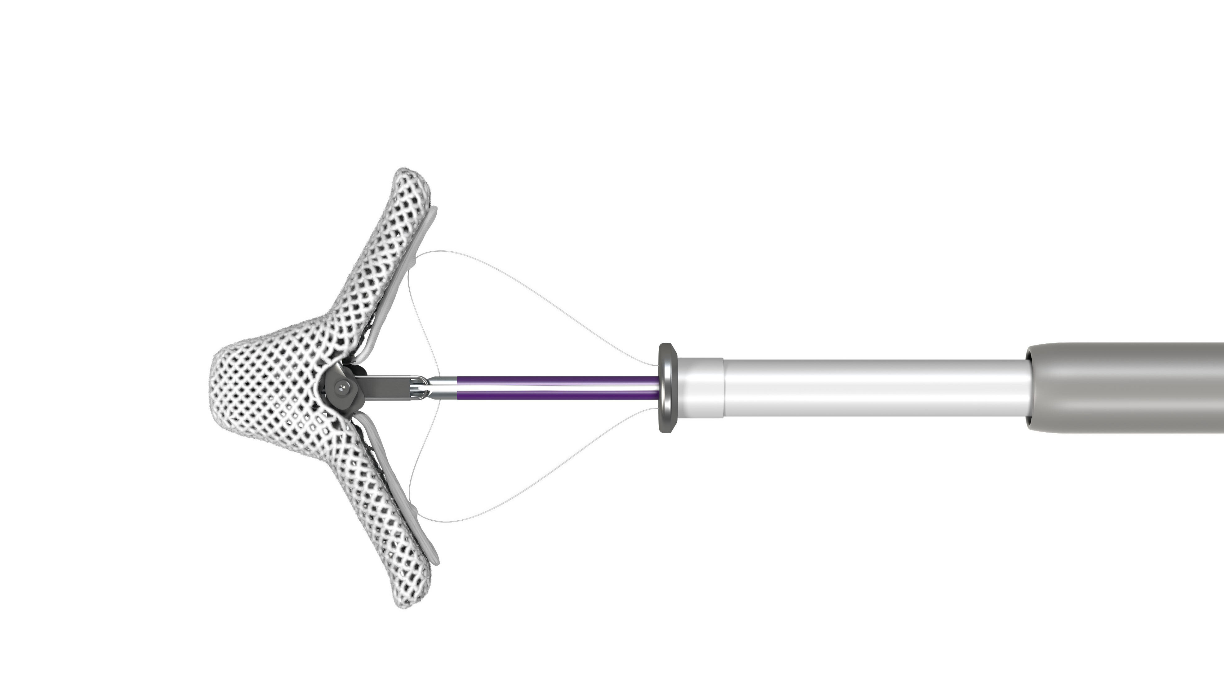
The MitraClip procedure
In the MitraClip procedure, a catheter is inserted via the inguinal vein into the right atrium and through the atrial septum into the left atrium. The clip is brought to the mitral leaflets via this catheter and the two leaflets are pulled together at certain points.
(Photo: Abbott)
The Structural Heart Interventions Programme (SHIP)
Every year, more than 1,500 interventional heart valve procedures are performed at the Deutsches Herzzentrum der Charité (DHZC). This makes the DHZC a world leader in this field.
Our cardiac surgery and cardiology teams have been working closely together for years in the treatment of heart valve patients. This collaboration will be further expanded and consolidated in the new unit for catheter-based heart valve interventions, the ‘Structural Heart Interventions Programme (SHIP)’.
In the Structural Heart Interventions Programme (SHIP) unit, we bundle the medical planning and implementation of all catheter-based treatments of heart valves in adults. The treatments carried out for heart valve diseases primarily include TAVI procedures to replace the aortic valve, but also catheter-based treatments for diseases of the mitral and tricuspid valves.
All information on the services and the interdisciplinary team of the ‘Structural Heart Interventions Programme (SHIP)’ can be found here.
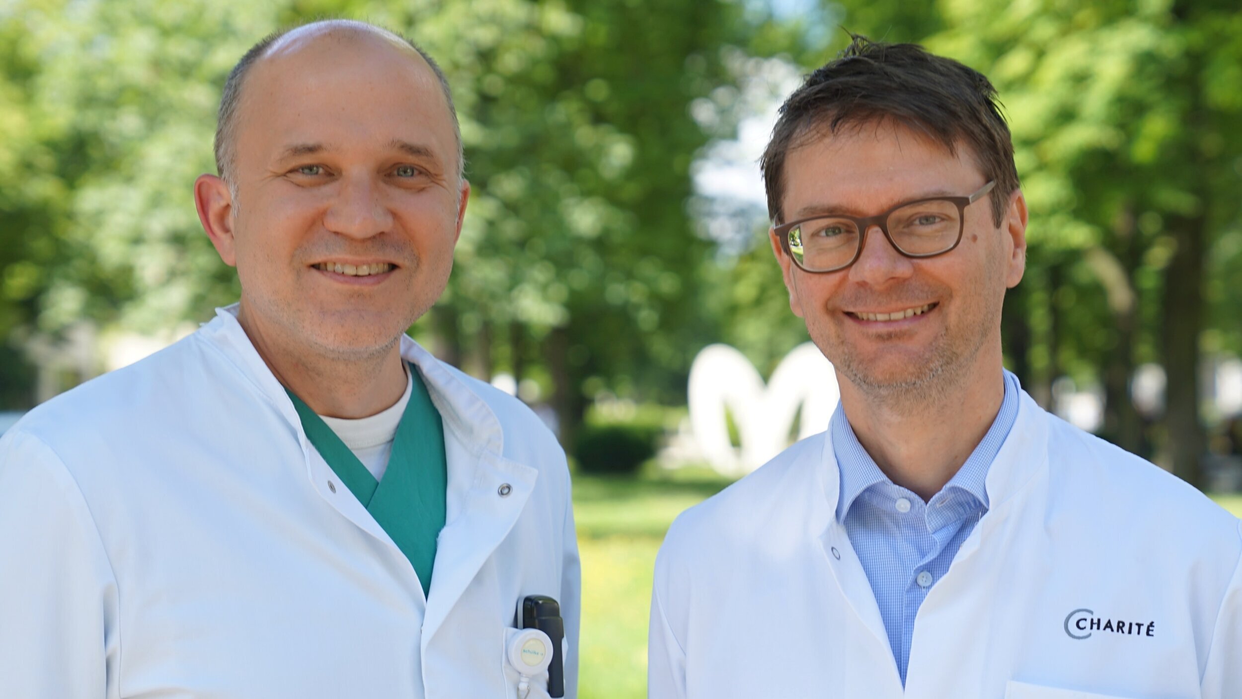
PD Dr Axel Unbehaun (left) and Prof. Dr Henryk Dreger (right) are the medical directors of the Structural Heart Intervention Programme (SHIP).
In this unit, patients are cared for by an interdisciplinary team - from admission to discharge and aftercare.
PD Dr Axel Unbehaun (left) and Prof. Dr Henryk Dreger (right) are the medical directors of the Structural Heart Intervention Programme (SHIP).
In this unit, patients are cared for by an interdisciplinary team - from admission to discharge and aftercare.
Certified mitral valve centre
The Deutsches Herzzentrum der Charité has been certified as a mitral valve centre by the German Society of Cardiology (DGK).
In the multi-stage assessment process, numerous documents must be provided to prove the successful diagnosis and treatment of mitral valve diseases over several years, such as case numbers, quality data, technical equipment as well as the experience of the specialists and their regular participation in specialist training courses.