Heart valves and their diseases
Each half of the heart has a semilunar valve and a sigmoid valve. The semilunar valves are located between the atrium and the ventricle and are called the mitral valve (left) and the tricuspid valve (right). The sigmoid valves are located between the ventricle and the outflow vessel and are called the aortic valve (left) and the pulmonary valve (right).
As valves in the heart, the four heart valves ensure that the blood only flows in one direction. In heart valve disease, one or more of the valves are either constricted, calcified or leaky.
The heart valves
The four heart valves: Aortic valve, mitral valve, tricuspid valve and pulmonary valve.
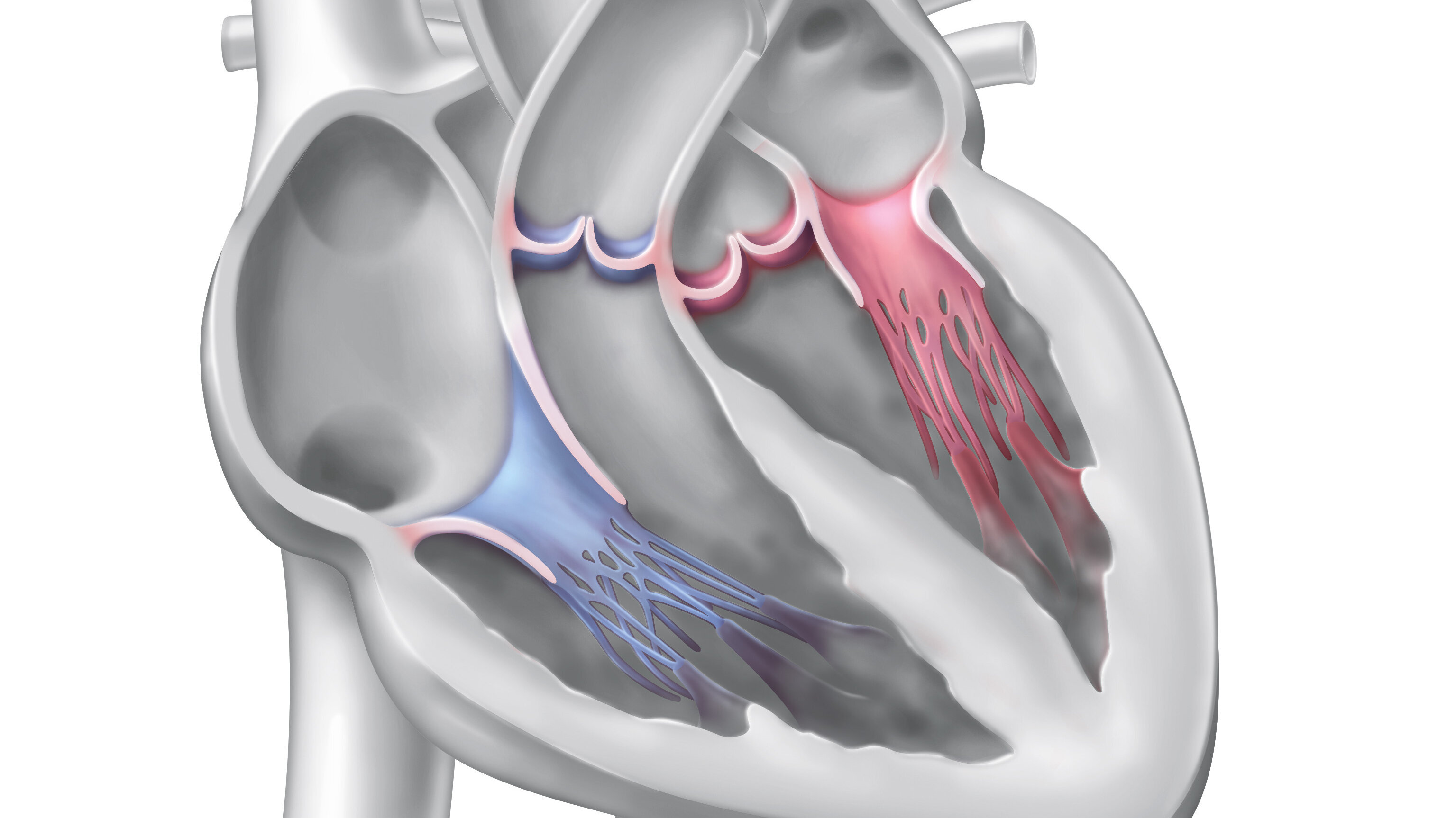
The heart valves
The four heart valves: Aortic valve, mitral valve, tricuspid valve and pulmonary valve.
Symptoms
Valvular heart disease can be congenital in rare cases, but it is much more common for it to be acquired – for example, through calcification in old age, inflammation or circulatory disorders. A malfunction of the valves can be caused either by restricted opening (valve stenosis) or insufficient closure (valve insufficiency) or a combination of the two. This disturbs the proper blood flow, causing the heart muscle to work harder.
If the function of the heart valves is only slightly restricted, this is usually not noticeable in everyday life. However, if the valve disease continues to progress, the heart muscle can no longer maintain an adequate blood flow: heart failure develops. This can lead to shortness of breath during physical exertion, coughing, dizziness, fainting, increased water retention in the legs or chest pain. But heart rhythm disorders can also be a consequence of heart valve disease.
Diagnosis
The earlier valve diseases and their accompanying symptoms are detected and controlled, the better they can be treated. At the DHZC, we offer the following diagnostic procedures:
- The typical noises associated with heart valve disease can already be detected by listening with a stethoscope.
- With the help of echocardiography, heart valve diseases and their effect on the heart muscle can be detected precisely and safely and their severity can be classified.
- Cardiac magnetic resonance imaging (MRI) is another state-of-the-art procedure that can be used if necessary to precisely assess the severity of the heart and valve disease.
- A cardiac catheter examination provides more precise information in the case of advanced disease and is helpful for further therapy planning.
- High-resolution computed tomography is used to analyse the geometry of the heart valves and the treatment paths to the heart valves. This enables us to treat these diseases gently and as minimally invasively as possible.
Therapy options
In the following, we will introduce you to the heart valves and their diseases in detail and explain which treatment methods we offer at DHZC. The type of therapy depends on the severity of the valve disease and the patient's complaints, as well as the chances and risks of the individual treatment options. All patients are analysed in an interdisciplinary team and receive detailed advice on the best possible and least risky strategy.
In the case of severe valve dysfunction, it is recommended to either repair or replace the affected valve. For this purpose, either minimally invasive procedures using catheters or surgical procedures are used. In the following, we will present the heart valves and their diseases in detail and explain which treatment methods we offer at DHZC.