Optical coherence tomography (OCT)
Optical coherence tomography (OCT) is an innovative, invasive imaging technique used in the diagnosis of coronary heart disease (CHD).
Vulnerable coronary plaque
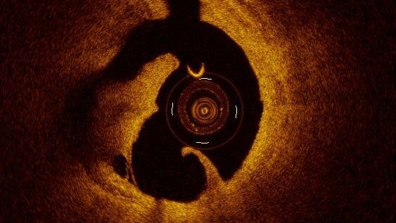
Plaque rupture
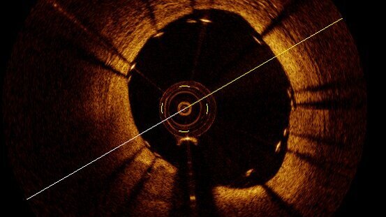
Check-up after stent implantation
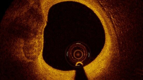
Vulnerable coronary plaque
Plaque rupture
Check-up after stent implantation
Method
In this procedure, a small catheter based on infrared light is used to display the coronary vessel wall in high resolution. This makes atherosclerotic changes in the coronary vessels, known as plaques, visible in terms of their extent and composition. This provides crucial additional information to coronary angiography, which forms the basis for the type and extent of interventional therapy in the cardiac catheter laboratory.
In addition, OCT provides a precise 3D real-time representation of the anatomy and pathophysiology of the coronary arteries. This optimises the planning, execution and success monitoring of coronary interventions (e.g. imaging of complex bifurcation stenoses, implantation of stents and bioresorbable vascular scaffolds).
Since 2015, the Department of Cardiology at the Charité's Benjamin Franklin campus has been one of the first centres worldwide to have a system for integrating 3-dimensional OCT image information and haemodynamic measurements of the ‘fractional coronary flow reserve’ (FFR) into coronary angiography. This makes it possible to perform optimal and very precise invasive diagnostics and to plan coronary interventions accurately.
Optical Coherence Tomography at the DHZC
The Department of Cardiology and Angiology at Campus Benjamin Franklin (CBF) offers the full range of invasive cardiological and angiological examinations and interventional therapies in modern interventional cardiology and angiology. Four state-of-the-art cardiac catheterisation laboratories with digital, biplane angiography systems are available for this purpose.
Appointments can be made through the cardiology secretariat of the outpatient clinic.