The electrocardiogram (ECG)
ECG stands for electrocardiogram and refers to the measurement of the electrical activity of the heart. The heartbeat, i.e. the rhythmic contraction of the heart muscle, is triggered by the so-called sinus node in the centre of the heart. From here, electrical impulses spread to all areas of the heart via branching pathways. This so-called cardiac action is recorded on the ECG in the form of curves - the famous ‘ECG waveform’. This provides the doctor with information about the state of health of the heart.
To carry out the ECG measurement, several ECG electrodes are attached to the patient's skin and connected via cables to the ECG device, which records the heart activity. The ECG is usually the first and quickest cardiological diagnostic option - a portable ECG is available in every ambulance.
A patient sits on an ergometer to perform an exercise ECG. You can see the sensors on his chest. He is wearing a mask.
© DHZC
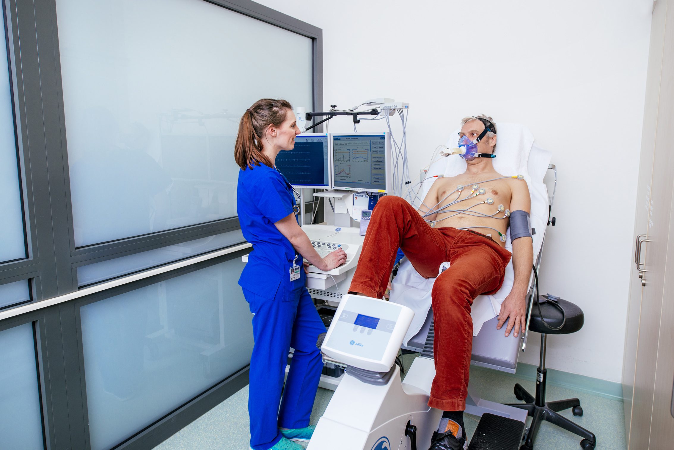
A patient sits on an ergometer to perform an exercise ECG. You can see the sensors on his chest. He is wearing a mask.
© DHZC
The exercise ECG
In some heart diseases, especially coronary heart disease, changes in the ECG often only become apparent during physical exertion. This is why the exercise ECG, known as ergometry, is a readily available, non-invasive examination method that can provide indications of a circulatory disorder in the heart. During the examination, the patient pedals on a cycle ergometer while the load gradually increases. The heart rate, blood pressure and ECG lead are recorded continuously. If there are indications of a circulatory disorder in the heart, there is usually an indication for further diagnostics, e.g. in the form of a cardiac catheterisation.
The ECG provides reliable information about the health of the heart and is usually the first and quickest cardiological diagnostic option.
© DHZC
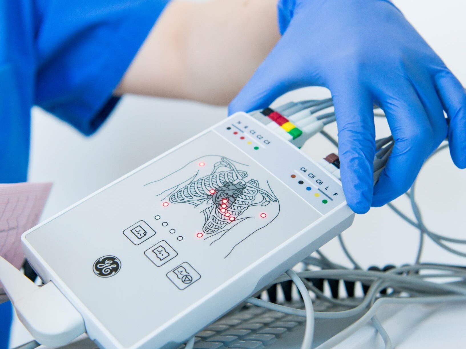
The ECG provides reliable information about the health of the heart and is usually the first and quickest cardiological diagnostic option.
© DHZC
Long-term ECG and loop recorder
Longer-lasting cardiac arrhythmias can be diagnosed with the help of a resting ECG. However, if they only occur occasionally, a long-term ECG measurement is useful. In this case, an ECG device worn by the patient on a belt records the electrocardiogram over a period of 24 to 72 hours.
For even less frequent arrhythmias that cannot be recorded with long-term measurements over three days, we offer recording with a ‘loop recorder’. A distinction is made between a portable external recorder and an implantable recorder:
- With an external loop recorder , the device is worn on a belt and the ECG is recorded via attached skin electrodes. The recording is triggered by the patient by pressing a button as soon as he or she recognises an arrhythmia. The wearing time is about 10 - 14 days.
- The implantable recorder is inserted under the skin near the chest. It can record the heart rhythm for up to three years. The device is then removed again in a simple procedure. Recorded cardiac arrhythmias can be easily read out by placing an interrogation device on the patient.