Computed tomography (CT)
Computed tomography (CT) is based on the use of X-rays and, like MRI, produces many cross-sectional images. The patient lies in a rotating X-ray tube. The computer calculates an overall three-dimensional image from many two-dimensional X-ray images. CT is the method of choice for aortic diseases in order to be able to make a precise diagnosis.
Compared to MRI, the examination only takes a few minutes and can therefore also be used in an emergency situation. At the DHZC, we use a modern dual-source CT scanner in which two X-ray tubes rotate around the body at the same time. This allows scans to be carried out in a matter of seconds and without ‘breath-holding’. Further information about our CT scanners can be found here.
Contrast agents are often used to better assess blood vessels (e.g. the aorta). This examination is called CT angiography.
The staff of the Cardiology Department in front of a computer tomograph. In the foreground Dr Natalia Solovyova.
© DHZC
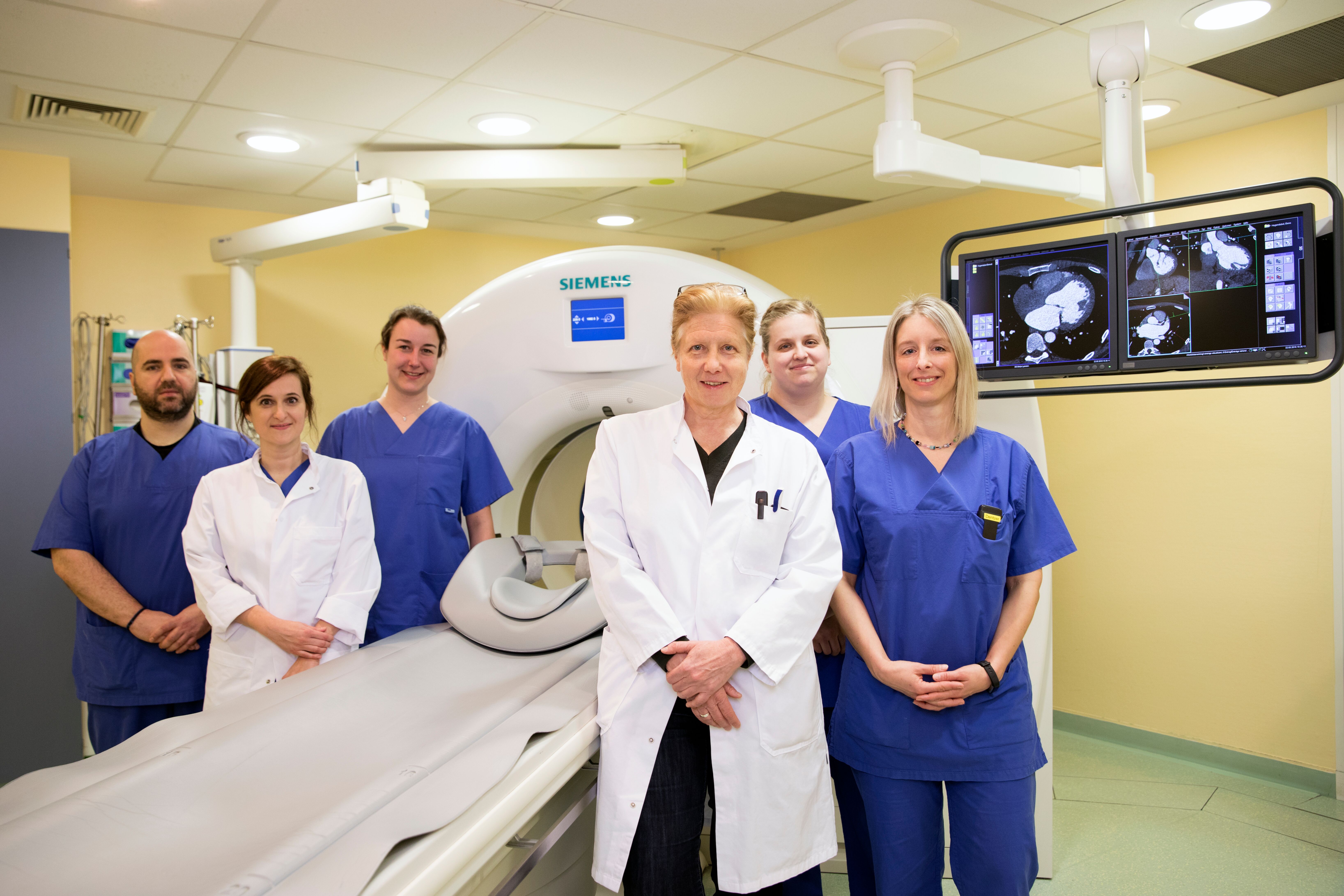
The staff of the Cardiology Department in front of a computer tomograph. In the foreground Dr Natalia Solovyova.
© DHZC
When is a CT scan necessary?
The cardiological societies recommend a CT scan for patients at low and medium risk of coronary heart disease (CHD). CT scans are also an alternative to invasive cardiac catheterisation for patients with known CHD and especially after ACVB surgery, or can be used to help decide whether a cardiac catheterisation is necessary. Computed tomography is not recommendedas a preventive examination.
Further areas of application of CT in our clinic:
- Diseases of the large (aorta) and small vessels (e.g. neck and leg vessels)
- before valve operations (TAVI)
- for metallic implants to determine the heart anatomy and function
- for follow-up monitoring after a heart transplant
- for patients with congenital heart defect
Positron emission tomography (PET-CT)
Positron emission tomography (PET-CT) uses radioactively labelled substances to measure metabolic activity in tissue. It is also a cross-sectional examination that can be combined with a CT scan. In cardiology, for example, PET-CT is used to assess the vitality of the heart muscle. In aortic surgery, inflammation of the aorta or a prosthesis infection can be visualised using radioactively labelled sugars. Inflamed tissue is more metabolically active and consumes more sugar, which is why a particularly large number of radioactively labelled substances accumulate in this area.