Electrophysiological examination (EPU)
Electrophysiological examination (EPU) is a special form of cardiac catheter examination. It is used to diagnose cardiac arrhythmias and to provide treatment tailored to the individual patient.
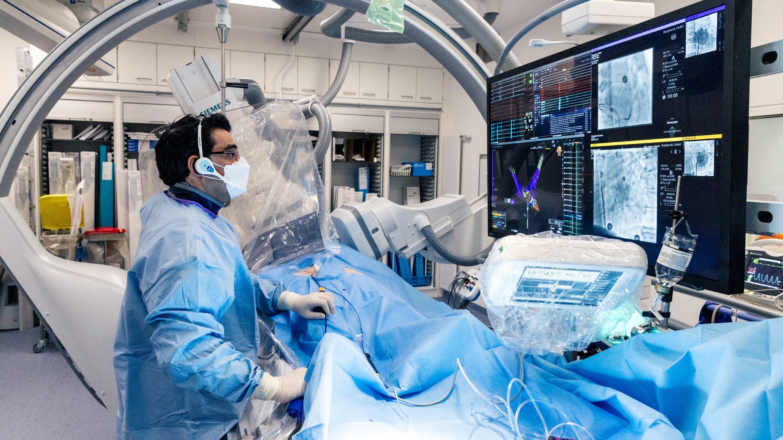
During an EPU, the cardiologist places several catheters fitted with electrodes in the heart. These allow electrical signals to be derived directly from the heart and can be combined with various visualisation techniques (3D visualisation, fluoroscopy using X-rays).
The aim of an electrophysiological examination (EPU) is to diagnose and treat cardiac arrhythmia. This examination is used to analyse the electrical impulses and processes in the heart in order to detect disturbances in the heart rhythm.
The aim of an electrophysiological examination (EPU) is to diagnose and treat cardiac arrhythmia. This examination is used to analyse the electrical impulses and processes in the heart in order to detect disturbances in the heart rhythm.
Reasons for realising an EPU
An EPU is performed in these cases, among others:
- For the diagnosis of cardiac arrhythmias: An EPU helps to identify the type, origin and cause of cardiac arrhythmias such as atrial fibrillation, ventricular tachycardia or other arrhythmias.
- To determine the treatment strategy: The examination can determine whether drug therapy, a pacemaker, a defibrillator or ablation is necessary. It also helps to better assess the risk of sudden cardiac death or other serious complications.
- Targeted ablation therapy: An EPU can lead directly to therapy by specifically obliterating the area causing the arrhythmia by means of an ablation procedure. This allows the normal heart rhythm to be restored. In this case, an EPU serves as preparation for ablation.
- For the evaluation of medication: The examination can be used to test the effectiveness of certain medications for the treatment of cardiac arrhythmias.
An electrophysiological examination is particularly helpful for patients who suffer from persistent or intermittent cardiac arrhythmias and helps with both diagnosis and targeted treatment to improve the patient's quality of life.
Process of an EPU
An electrophysiological examination (EPU) is carried out in several steps and is usually performed under local anaesthetic and sometimes under light sedation. The procedure is usually as follows:
Preparation:
- The patient is prepared for the procedure in a sterile examination area in a special cardiac catheterisation laboratory.
- In most cases, a mild sedative is administered and the relevant area (the right and/or left groin) is shaved and sterilised.
- The patient is connected to monitoring equipment to continuously check heart rhythm, blood pressure and oxygen levels.
Procedure site and access:
- The doctor anaesthetises the puncture site in the groin to create access for the electrode catheters.
- Flexible, thin electrode catheters are inserted via a vein or artery and carefully advanced into the heart under X-ray control.
Measurement and analysis:
- As soon as the electrodes are positioned in the heart, electrical impulses are emitted to specifically provoke cardiac arrhythmia and check the electrical conductivity in the heart.
- The heart's reaction is recorded and analysed to determine whether and where there is a disturbance, for example in the atria or ventricles.
- During this phase, the doctor can stimulate different areas of the heart to identify possible arrhythmias and better understand their triggers.
Diagnosis or treatment decision:
- Based on the results of the examination, the diagnosis and appropriate therapy are determined on an individual basis.
- EPU is often combined directly with catheter ablation to eliminate the cause of the arrhythmia. During an ablation, the tissue causing the cardiac arrhythmia is specifically sclerosed.
Completion of the examination:
- Once the examination is complete, the electrode catheters are carefully removed and the puncture site is treated.
- The patient must then remain in hospital for a few hours or up to a day for observation to rule out bleeding or other possible complications.
Aftercare:
- After the EPU, the patient should take it easy for a few days and avoid physical exertion.
- In the case of an ablation that often follows, a follow-up examination is often scheduled to ensure that the cardiac arrhythmia does not recur.
Duration:
A simple EPU takes about 30 minutes. However, depending on the complexity and origin of the cardiac arrhythmia, this duration may be extended accordingly.
Possible complications:
The examination is considered very safe, but complications can occur in rare cases. These include bleeding and haematomas at the puncture site, blood clots, injuries to the heart wall, infections, allergic reactions and cardiac arrhythmias.
The EPU provokes arrhythmias in order to identify their cause. However, sometimes these can also occur briefly after the procedure. However, they usually normalise on their own or can be treated with medication.
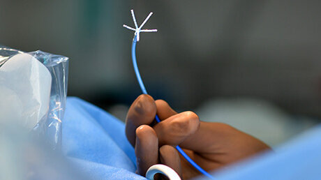
In a catheter ablation procedure, probes are inserted into the heart, which can be used to sclerose heart muscle tissue at the sites that are causing the arrhythmia.
(Image: DHZC)
In a catheter ablation procedure, probes are inserted into the heart, which can be used to sclerose heart muscle tissue at the sites that are causing the arrhythmia.
(Image: DHZC)
Treatment at the DHZC
Electrophysiological examinations often serve as preparation for subsequent catheter ablation. At the DHZC, they are carried out at all cardiology clinics at our locations in Berlin-Steglitz, Berlin-Mitte and Berlin-Wedding. At the DHZC, we have eight electrophysiological catheterisation laboratories with state-of-the-art equipment. We carry out more than 1,400 electrophysiological examinations and catheter ablations every year. We also advise around 4,000 patients a year in the rhythm consultation for further clarification of cardiac arrhythmias.
Our services for you
As a rule, you will receive a referral from your general practitioner if you are to be treated at our clinic for a cardiac arrhythmia. For an inpatient stay, you will need a referral slip or a prescription for hospital treatment. You will receive this from your general practitioner or specialist. You can find more information about an inpatient stay at the DHZC here.
In addition to the diagnosis and treatment of cardiac arrhythmias using an EPU, we also offer patients the opportunity to obtain a second opinion before the planned procedure. For example, if you receive a referral for EPU from your doctor, the DHZC offers to review this case.
For inpatients:
Patient management
T: +49 30 450 513 747
For outpatients:
Cardiological outpatient clinic
T: +49 30 450 513 717
For inpatients:
Patient management
T: +49 30 450 513 021
For outpatients:
Cardiological outpatient clinic
T: +49 30 450 513 150
For inpatients:
T: +49 30 450 565 400 (after the announcement: 1)
For outpatients:
T: +49 30 450 565 400 (after the announcement: 2)
Questions and answers
How long does the examination take?
A simple EPU takes about 30 minutes. However, depending on the complexity and origin of the cardiac arrhythmia, this time may be longer.
When can I go home after the examination?
The patient must remain in hospital for a few hours or up to a day for observation after the examination in order to rule out bleeding or other possible complications.
How many examinations need to be performed?
For most patients, only one EPU is required to make a clear diagnosis of the cardiac arrhythmia and possibly carry out treatment (e.g. ablation) straight away. However, in the case of more complicated or rare arrhythmias, several examinations may be necessary to find and treat the exact cause.
Is an EPU performed under anaesthetic?
An electrophysiological examination (EPU) is usually performed under local anaesthetic and sometimes under light sedation.
Authors


Verena Tscholl is a senior physician in the Rhythmology Department at the DHZC Department of Cardiology, Angiology and Intensive Care Medicine at Campus Charité Mitte. She is a specialist in internal medicine and cardiology with additional qualifications in cardiac pacing and as an ‘Electrophysiology Specialist I’ from the EHRA (‘European Heart Rhythm Association’).