Cardiovascular Imaging at the DHZC
Campus Virchow-Klinikum
Imaging procedures such as echocardiography, computerised tomography (CT) and magnetic resonance imaging (MRI) allow a view of the heart from the outside. Cardiac imaging is indispensable for recognising cardiovascular diseases at an early stage and treating them appropriately. At the DHZC, we use state-of-the-art, high-performance imaging techniques to provide our patients with the best possible care.
The cardiovascular imaging team at the DHZC, Campus Virchow-Klinikum comprises a total of 19 people. It is interdisciplinary and consists of colleagues from the
- Department for Cardiology, Angiology and Intensive Care Medicine at Campus Virchow-Klinikum,
- Department of Cardiothoracic and Vascular Surgery,
- Department of Congenital Heart Desease – Pediatric Cardiology,
- der Clinic for Radiology (des Charité Centrums CC06).
The department reports to Prof. Dr med. Volkmar Falk as Medical Director of the DHZC. He comprises the MRI department and the CT/X-ray department.
Prof. Dr Sebastian Kelle (cardiologist, senior physician at the Clinic for Cardiology, Angiology and Intensive Care Medicine at the Virchow-Klinikum campus) will head the MRI department for adult patients and Prof. Dr Titus Kühne (paediatric cardiologist at the DHZC and head of the Institute for Cardiovascular Computer-Assisted Medicine ICM at the DHZC) for children.
Dr Boris Gorodetski, radiologist and specialist at the Charité Clinic for Radiology (with the paediatric radiology department), is in charge of the CT/X-ray department.
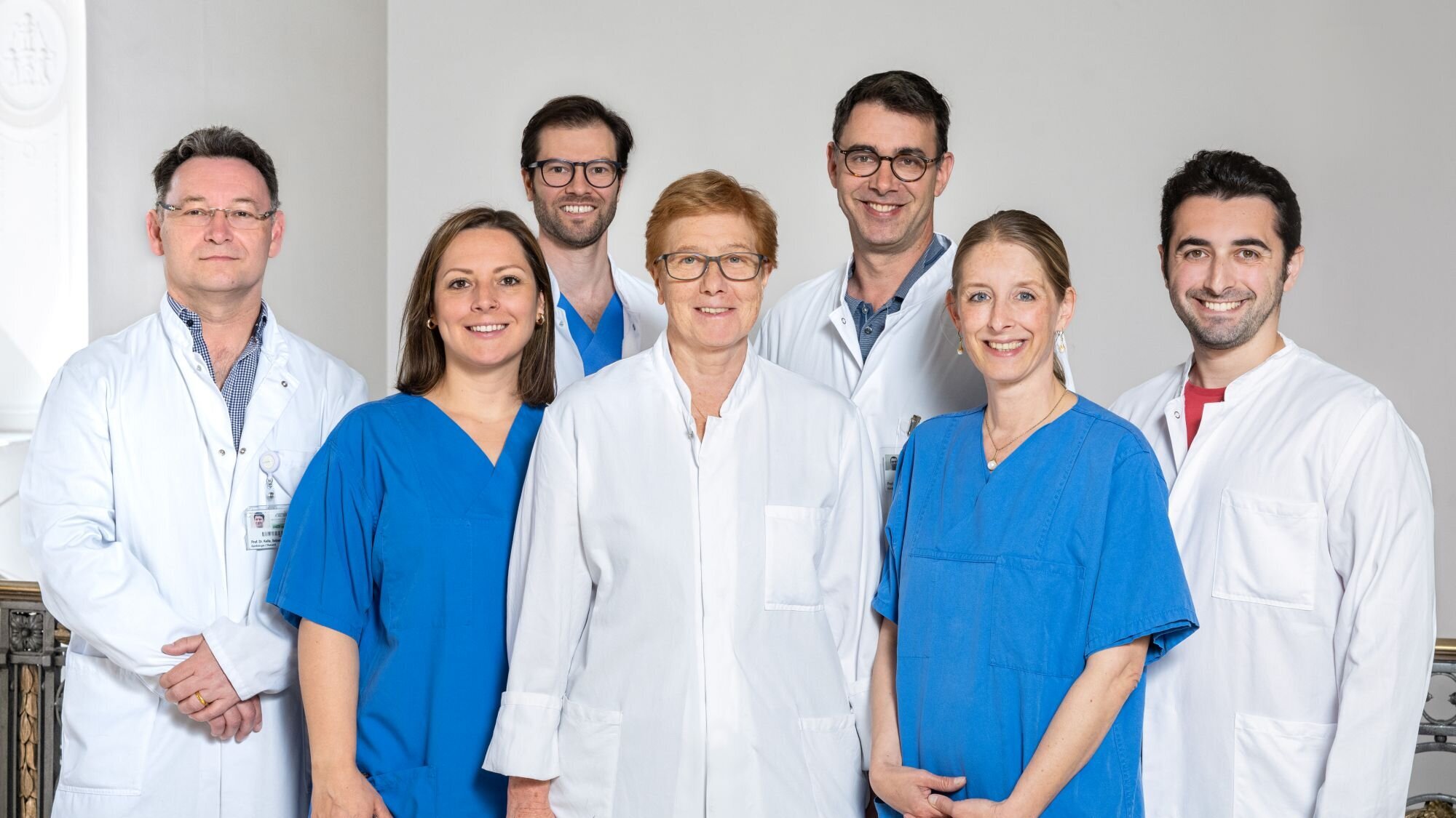
The cardiovascular imaging team at the DHZC, Campus Virchow-Klinikum:
Prof. Dr Sebastian Kelle, Annika Thillmann, Dr Patrick Doeblin, Dr Natalia Solowjowa, Prof. Dr Titus Kühne, Janina Dentzer and Dr Boris Gorodetski (from left to right).
The cardiovascular imaging team at the DHZC, Campus Virchow-Klinikum:
Prof. Dr Sebastian Kelle, Annika Thillmann, Dr Patrick Doeblin, Dr Natalia Solowjowa, Prof. Dr Titus Kühne, Janina Dentzer and Dr Boris Gorodetski (from left to right).
How we work: Our high-tech devices
Whether computed tomography (CT) or magnetic resonance imaging (MRI) - the imaging team at the DHZC only uses the latest generation of equipment, which is equipped with all available functions. This high-tech equipment makes the DHZC a global pioneer in the field of cardiovascular imaging.
Computertomographie (CT)
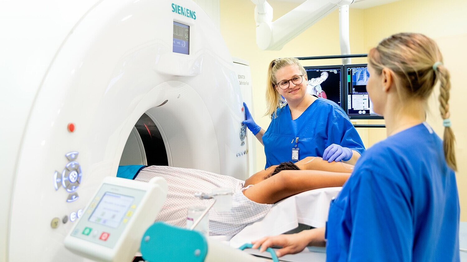
A computed tomography (CT) scanner can be used to visualise complex valvular heart disease or the small vessels of the heart with high spatial resolution. This makes it possible, for example, to quickly determine whether a patient is suffering from coronary heart disease. At the DHZC, we use a modern computer tomograph that guarantees optimum quality - and all this with a minimal radiation dose.
The heart presents every CT scanner with real challenges, as the heart rate and heart rhythm are different for every patient. The high-performance device available at the DHZC delivers fast and precise scans regardless of the heart rate and also makes it possible to reliably identify diseases. This allows the patient's treatment to be customised in a targeted manner.
Computed tomography (CT) can be used to visualise complex heart valve diseases or the small vessels of the heart with high spatial resolution.
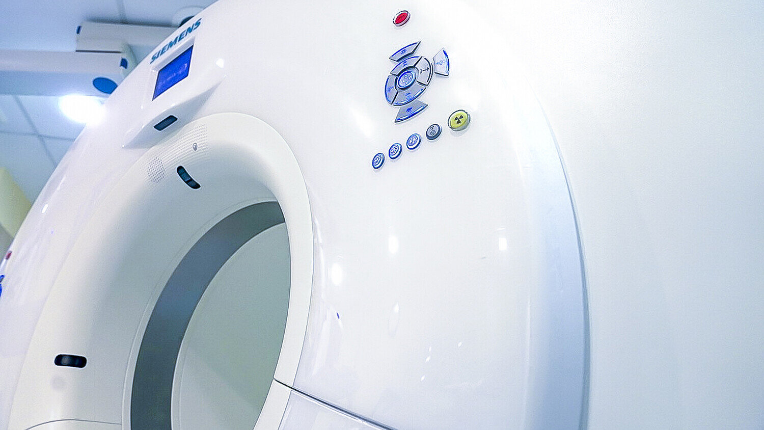
Computed tomography (CT) can be used to visualise complex heart valve diseases or the small vessels of the heart with high spatial resolution.
Magnetic resonance imaging (MRI)
At the DHZC, we have been using cardiovascular magnetic resonance imaging (MRI) for almost 30 years. It ensures that various heart diseases are recognised quickly and reliably. We work with two devices of the latest generation: a 1.5T and a 3T scanner. Both devices have the largest possible tube diameter of 70 centimetres, which makes the examinations much more comfortable and pleasant for patients.
Our magnetic resonance imaging scanners are faster, more energy-efficient and more comfortable than older models and also make it possible to examine patients with implants such as pacemakers and defibrillators without any problems. The automatic examination control relieves the MTRs and frees up time to concentrate on planning the examination. The devices' latest functions also ensure an improved workflow, especially for demanding cardiac examinations.
Our magnetic resonance tomographs are fast, energy-saving and comfortable. They enable us to easily examine patients with implants such as pacemakers and defibrillators.
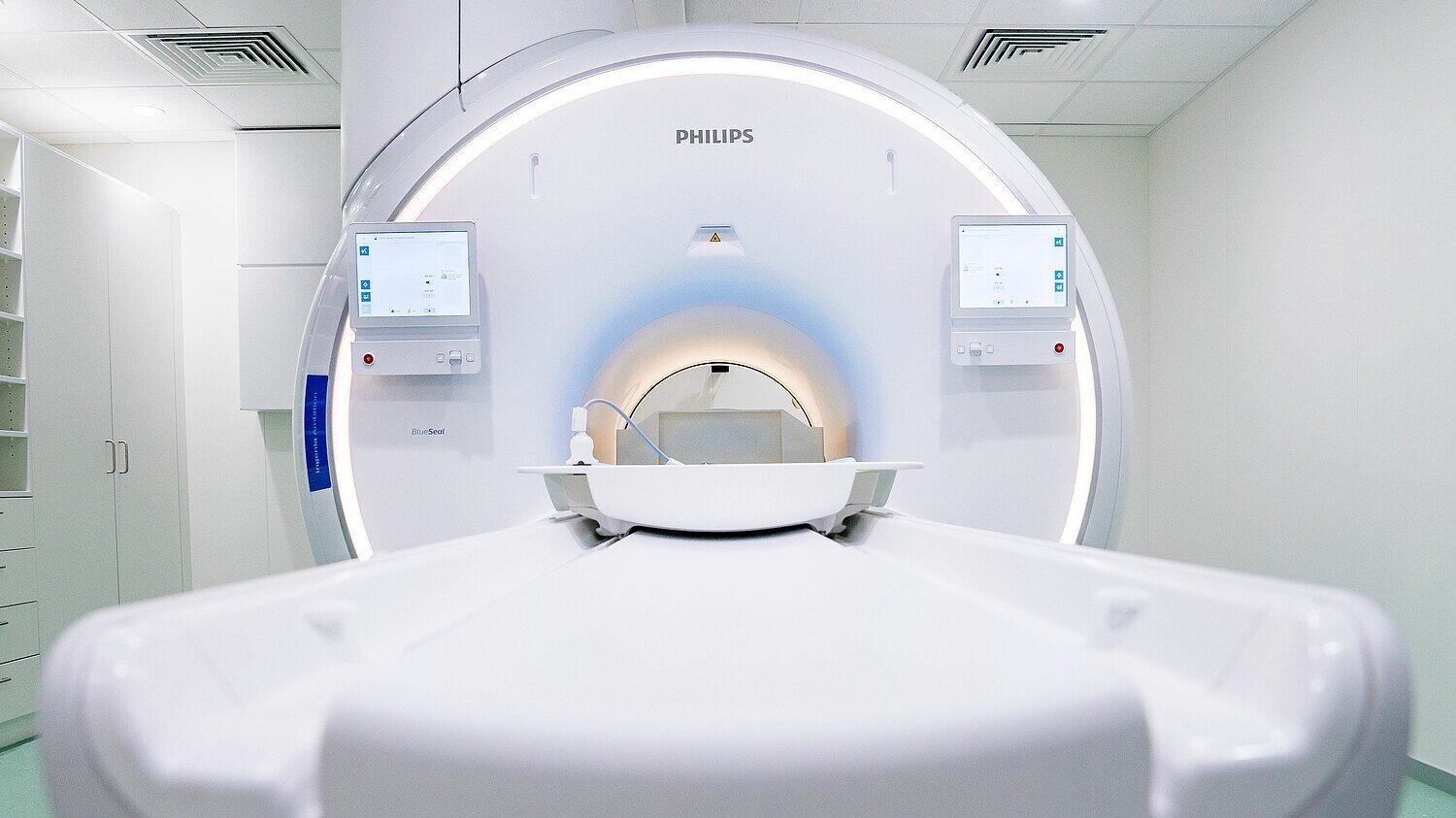
Our magnetic resonance tomographs are fast, energy-saving and comfortable. They enable us to easily examine patients with implants such as pacemakers and defibrillators.
X-ray
The Digital Diagnost C 90 upright X-ray unit enables convenient examination procedures and shorter waiting times. Innovative tools ensure efficient workflows. The live camera, flexible room configurations and examination automation techniques make examinations particularly pleasant for patients. UNIQUE 2 image processing quickly generates digital images of outstanding quality.
Seeing through the engine of life
Katharina works as a medical technologist for radiology (MTR) in the imaging team at the Virchow-Klinikum campus. In an in-depth interview, she explains what makes her work at the DHZC so exciting and how varied her tasks are.
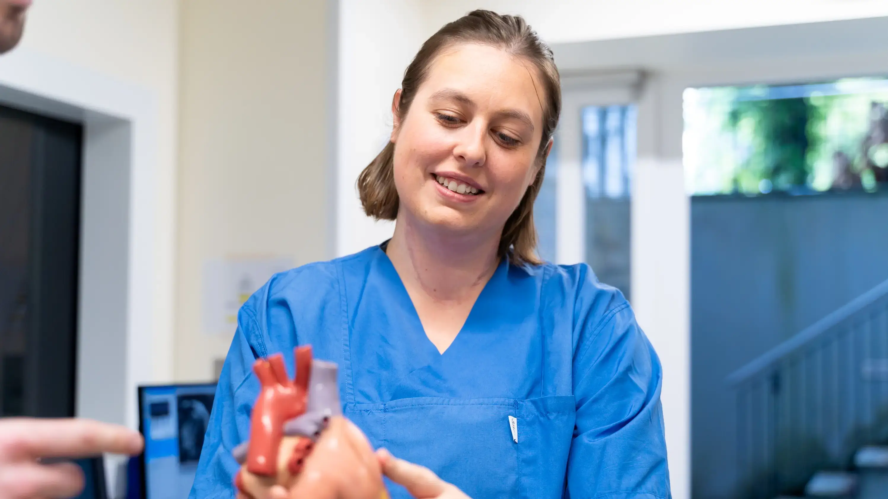
Seeing through the engine of life
Katharina works as a medical technologist for radiology (MTR) in the imaging team at the Virchow-Klinikum campus. In an in-depth interview, she explains what makes her work at the DHZC so exciting and how varied her tasks are.
We pass on our knowledge
20 years ago, we founded the CMR Academy to train external radiologists, cardiologists and nuclear medicine specialists in MRI diagnostics. The German Society of Cardiology (DGK) has certified the DHZC as a training centre for cardiac MRI and cardiac CT examinations up to the highest level (Level III) as one of only a few centres in the whole of Germany. Philips employees also receive further training at the DHZC. Several times a year, training courses are also held for external MTRs to qualify them to perform cardiac MRI examinations in collaboration with Philips.
We are looking for nice colleagues and team members!
At the DHZC, we offer MTRs and MFAs with an X-ray licence state-of-the-art technologies, flat hierarchies and a collegial team.
We are looking for beginners and experienced professionals to join the team and provide insight. A comprehensive induction concept ensures a good start in the team.
At the DHZC, we offer MTRs and MFAs with an X-ray licence state-of-the-art technologies, flat hierarchies and a collegial team.
We are looking for beginners and experienced professionals to join the team and provide insight. A comprehensive induction concept ensures a good start in the team.