Aortic valve stenosis is the most common heart valve defect requiring treatment. The aortic valve is narrowed and obstructs the flow of blood from the left ventricle into the aorta. In most cases, this disease is acquired. As the disease cannot be fundamentally improved and treated with medication, a narrowed aortic valve must be replaced. At the DHZC, we offer various procedures and heart valve prostheses.
Aortic valve insufficiency occurs less frequently. In this case, the aortic valve no longer closes completely. Mild forms of the disease do not need to be treated. An operation is only necessary if the function of the aortic valve is severely impaired. The valve can either be reconstructed or replaced. In some cases it is also necessary to treat the aorta. We perform all procedures at the DHZC.
Aortic valve stenosis
If the pockets of the aortic valve no longer open sufficiently for the blood flow, this is known as aortic valve stenosis (narrowing) - the most common heart valve defect. In this case, the aortic valve is narrowed and obstructs the flow of blood from the left ventricle into the aorta.
Symptoms
The following symptoms can occur with aortic valve stenosis:
- Shortness of breath
- Feeling of pressure on the chest
- dizziness
- Reduced performance
- Fainting spells
These symptoms usually occur during physical exertion, for example when climbing stairs. If aortic valve stenosis is not treated, the symptoms can also occur at rest over time and life expectancy can be dramatically reduced without treatment.
Reasons
In most cases, aortic valve stenosis is acquired, most frequently due to wear and tear processes such as calcification, thickening and hardening of the tissue in old age. Rheumatic fever or inflammation of the inner lining of the heart (endocarditis) can also lead to scarring of the aortic valve and cause aortic valve stenosis. Three to five per cent of people over the age of 75 suffer from aortic valve stenosis.
Congenital aortic valve stenosis is much rarer and leads to symptoms at a younger age.
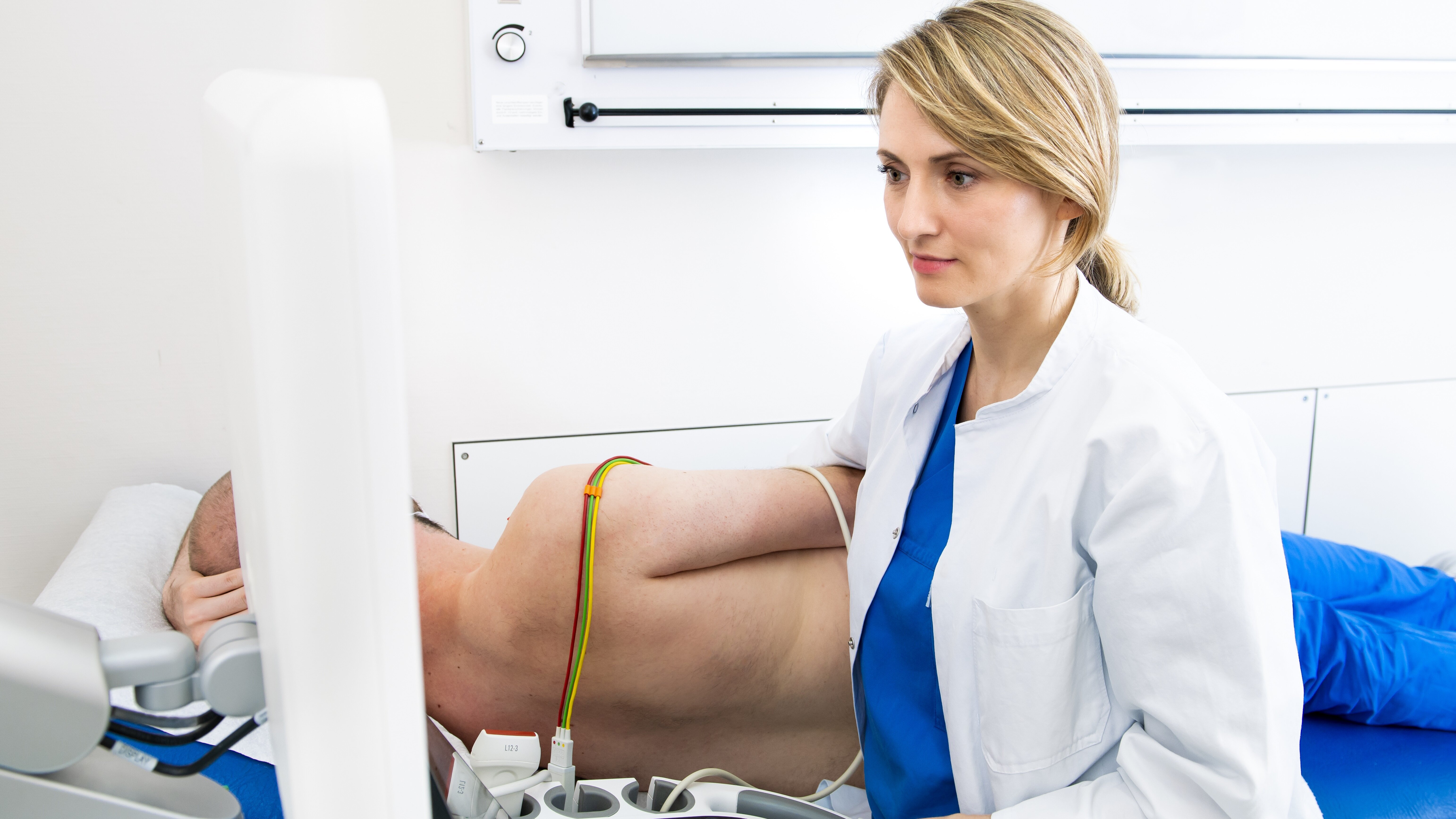
Echocardiography
Ultrasound waves are used to examine the heart ‘live’ and continuously.
The transducer receives the reflected waves and converts them into electrical impulses that are displayed on a screen.
(Image: DHZC/Külker)
Echocardiography
Ultrasound waves are used to examine the heart ‘live’ and continuously.
The transducer receives the reflected waves and converts them into electrical impulses that are displayed on a screen.
(Image: DHZC/Külker)
Diagnosis
To diagnose aortic valve stenosis, we offer the following examination methods at the DHZC:
- Echocardiography, the ultrasound examination of the heart
- Electrocardiogram (ECG), the measurement of the electrical activity of the heart
- Cardiac catheterisation
- Magnetic resonance imaging if necessary
- High-resolution computer tomography if necessary
Detailed information on the individual methods can be found on the respective pages.
Therapy
There is no drug therapy for aortic valve stenosis that significantly improves the prognosis of the disease. A severely narrowed aortic valve must be replaced.
A basic distinction must be made between surgical aortic valve replacement and catheter-assisted aortic valve replacement (TAVI). Replacement using the TAVI procedure is preferablyperformed without surgery and without a general anaesthetic, using only a puncture in the inguinal artery. For surgical replacement, minimally invasive techniques are also available as an alternative to complete transection of the sternum, in which the heart valve is replaced via an approx. 5 cm long incision in the chest.
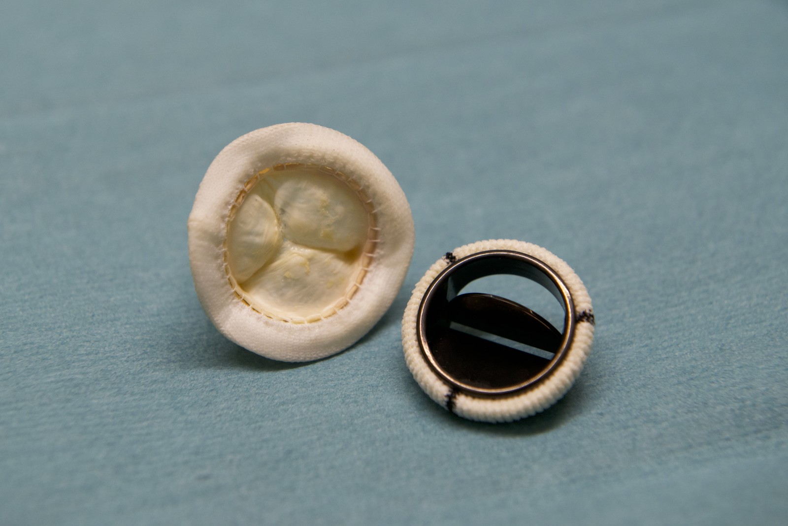
Heart valve prosthesis
A biological heart valve prosthesis on the left and a mechanical heart valve prosthesis on the right.
Heart valve prosthesis
A biological heart valve prosthesis on the left and a mechanical heart valve prosthesis on the right.
Deciding which procedure offers the best possible and most sustainable result requires a detailed analysis of the individual medical history and the findings of the imaging obtained. Our patients are advised on the basis of this assessment and the constantly updated findings of medical science and the recommendations of the professional associations.
The treatment is determined together with you. Participation in studies is also examined and, if suitable, offered as a possible alternative.