Magnetic resonance imaging (MRI)
Magnetic resonance imaging (MRI) uses a strong magnetic field to generate many cross-sectional images. Soft tissue and nerve tissue can be assessed particularly well in this way. The examination method shows the exact anatomy and function of heart defects and helps doctors to make important decisions when treating patients.
Magnetic resonance imaging shows the exact anatomy and function of heart defects and thus helps to determine the appropriate therapy for the patient.
© DHZC/Külker
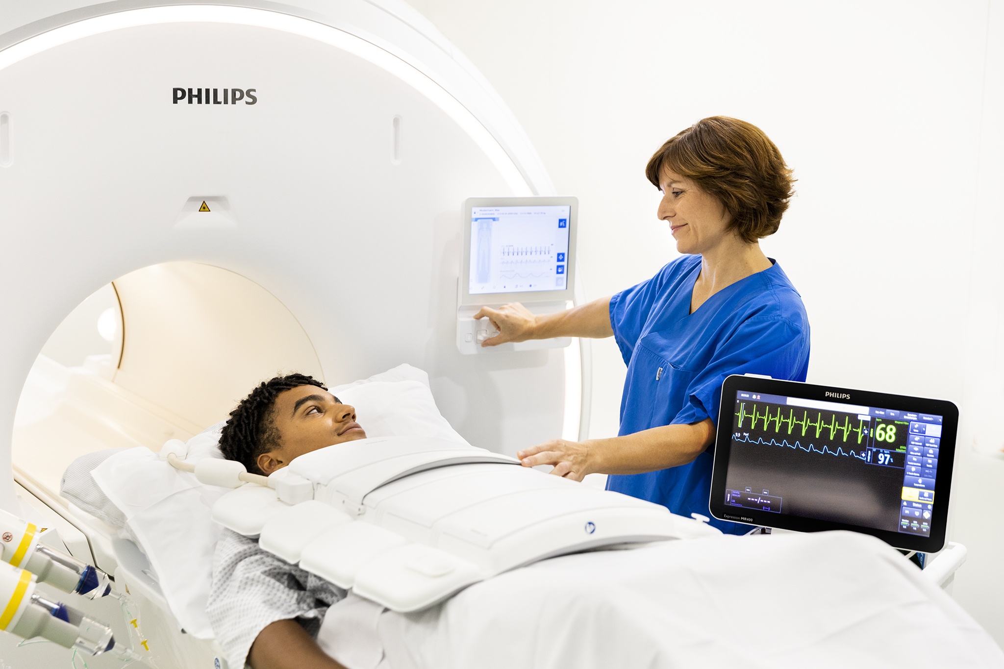
Magnetic resonance imaging shows the exact anatomy and function of heart defects and thus helps to determine the appropriate therapy for the patient.
© DHZC/Külker
Advantages of MRI
The great advantage of MRI is that it provides information on the anatomy, function and condition of the tissue in a single examination. MRI is often superior to other imaging procedures because it allows precise and - unlike a CT scan - radiation-free measurements.
However, as the examination takes a relatively long time, it cannot be carried out in an acute situation. Furthermore, MRI is not possible for patients with metal in their bodies, for example due to various prostheses or pacemakers, because of the strong magnetic field.
Areas of application
After a heart attack, parts of the heart muscle no longer work sufficiently. With the help of MRI, it is possible to distinguish which area is dead and which is still vital - regardless of when the heart attack occurred. This can be particularly important before bypass operations or catheter-based interventions.
For patients with suspected circulatory disorders of the heart muscle, an MRI scan is useful. During the examination, medication can be administered to cause a temporary strain on the heart. This allows the doctor to recognise how well the heart muscle is supplied with blood and which areas of the heart are not sufficiently supplied with blood under stress (adenosine stress MRI or dobutamine stress MRI). The performance of this so-called stress MRI is very safe and has a very high diagnostic accuracy.
Myocarditis is a disease that can lead to permanent heart failure and sudden cardiac death. As diagnosis with procedures such as ECG or echocardiography can be difficult, the use of MRI is often useful: inflammation of the heart muscle and pericardium can be visualised directly in MRI.
If a congenital heart muscle disease (cardiomyopathy) is suspected, MRI provides valuable additional information for making the correct diagnosis and as part of follow-up checks. MRI is regularly used to visualise the blood vessels (angiography) in the entire cardiovascular system.
An MRI examination enables precise and radiation-free measurements.
© DHZC
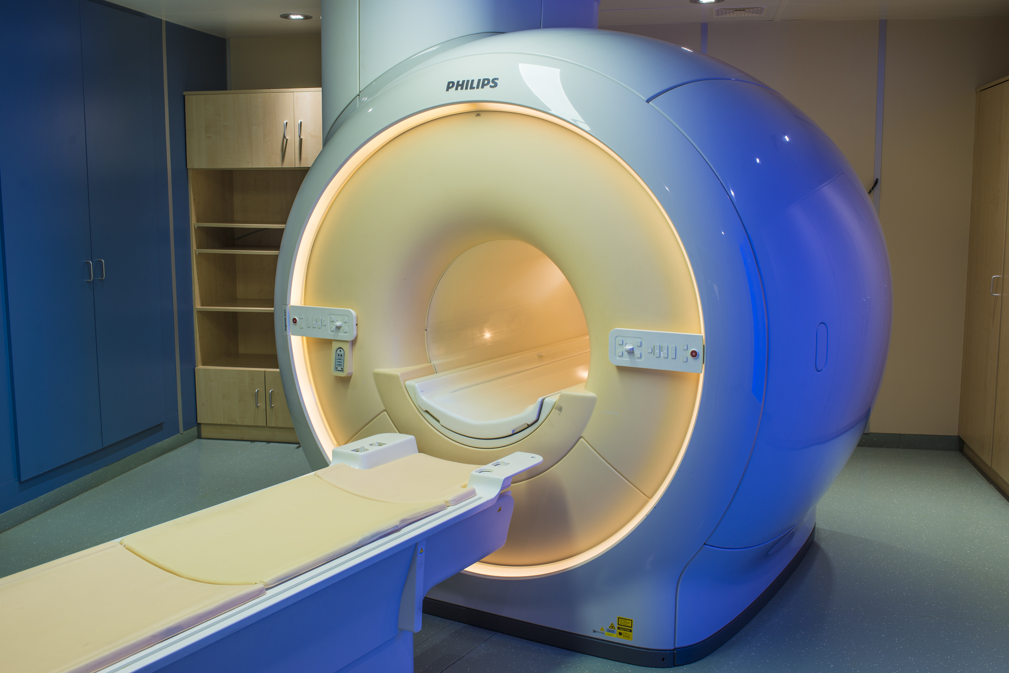
An MRI examination enables precise and radiation-free measurements.
© DHZC
MRT-Diagnostik am DHZC
Am Deutschen Herzzentrum der Charité setzen wir die MRT bereits seit 1996 ein. Unser Herzzentrum spielte von Anfang an eine führende Rolle bei der Entwicklung dieser diagnostischen Methode. 2001 wurde die CMR-Akademie gegründet, um unser Fachwissen in individuellen Kursen an Radiolog:innen, Kardiolog:innen und Nuklearmediziner:innen aus aller Welt weiterzugeben.
Als eines von wenigen Zentren bundesweit wurde unsere Klinik 2015 von der deutschen Gesellschaft für Kardiologie (DGK) als Ausbildungszentrum für kardiale MRT-Untersuchungen zertifiziert.