Echocardiography
Echocardiography is an ultrasound examination of the heart. Doctors also refer to it as a ‘cardiac echo’. Ultrasound waves are emitted via a so-called transducer and either ‘swallowed’ or reflected back by the body tissue. The transducer receives the reflected waves and converts them into electrical impulses that are displayed on a screen.
This happens ‘live’ so that the cardiologist can continuously examine the heart with the ultrasound.
Transthoracic echocardiography (TTE)
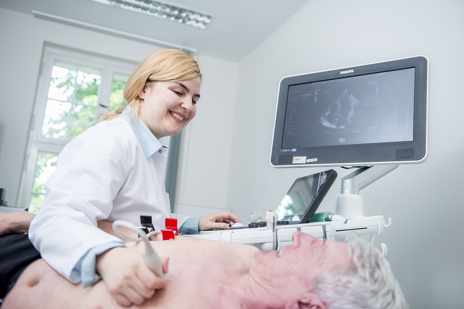
During transthoracic echocardiography, the doctor guides the ultrasound probe, which is about the size of a mobile phone, over the chest wall and aligns it differently in order to obtain as accurate an image as possible of the heart structures on the monitor. TTE is used as an initial guide to assess, for example, heart size, wall thickening, pumping and valve function. Doppler sonography and colour Doppler sonography provide additional information on the direction, speed and strength of blood flow in the vessels. Sonography is used, for example, to diagnose acute events such as a heart attack and is also suitable for monitoring the course of the disease.
While conventional echocardiography devices only provide two-dimensional images, we also use modern 3D echocardiography at the Deutsches Herzzentrum der Charité, which provides a three-dimensional image of the heart. Many millimetre-thin two-dimensional ultrasound images are taken in quick succession or simultaneously and displayed as a spatial structure. This means that in many cases, further transoesophageal echocardiography is not required for diagnosis.
TTE provides the most accurate image possible of the heart structures.
© DHZC
The ‘swallow echo’ is performed, for example, to exclude blood clots, for the early diagnosis of endocarditis or to examine the heart valves.
© DHZC
TTE provides the most accurate image possible of the heart structures.
© DHZC
The ‘swallow echo’ is performed, for example, to exclude blood clots, for the early diagnosis of endocarditis or to examine the heart valves.
© DHZC
Transoesophageal echocardiography (TEE)
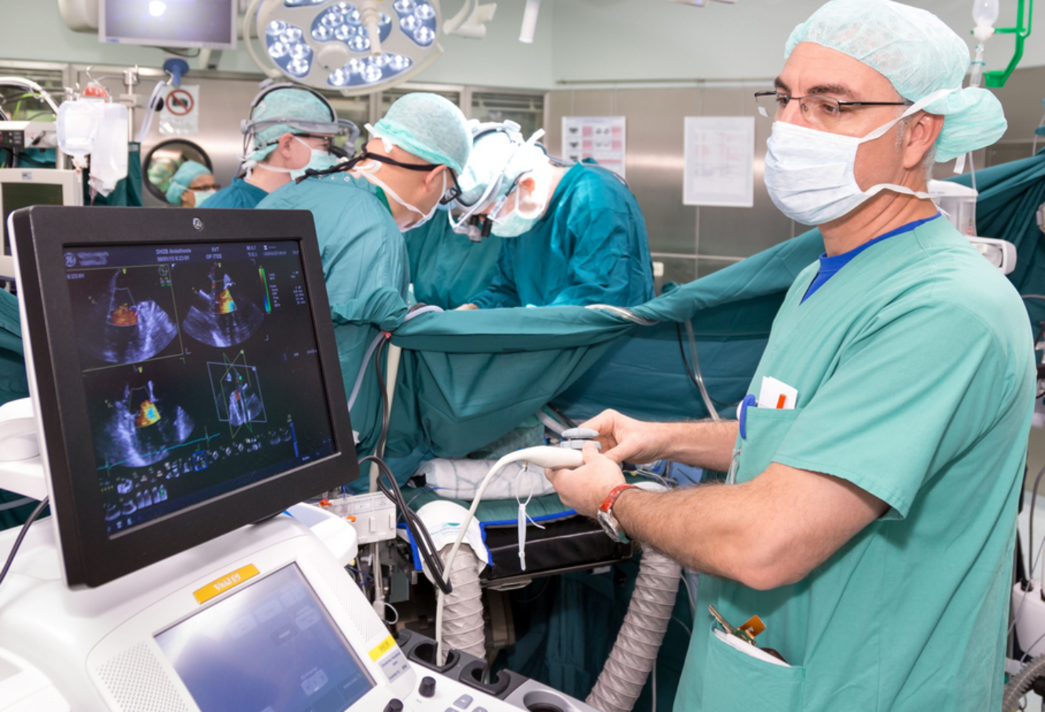
Transoesophageal echocardiography (TEE) is performed via the oesophagus and is therefore also known as ‘swallowing echo’. The patient is first given sedative medication. A very thin transducer is then inserted into the oesophagus. The advantage of this method is that the transducer is located below the sternum and close to the heart and therefore provides better images. Parts of the heart located deep in the chest can also be visualised.
A ‘swallow echo’ is performed, for example, to exclude blood clots, for the early diagnosis of endocarditis or to examine the heart valves. The Deutsches Herzzentrum der Charité is a certified training centre for this procedure.
Stress echocardiography
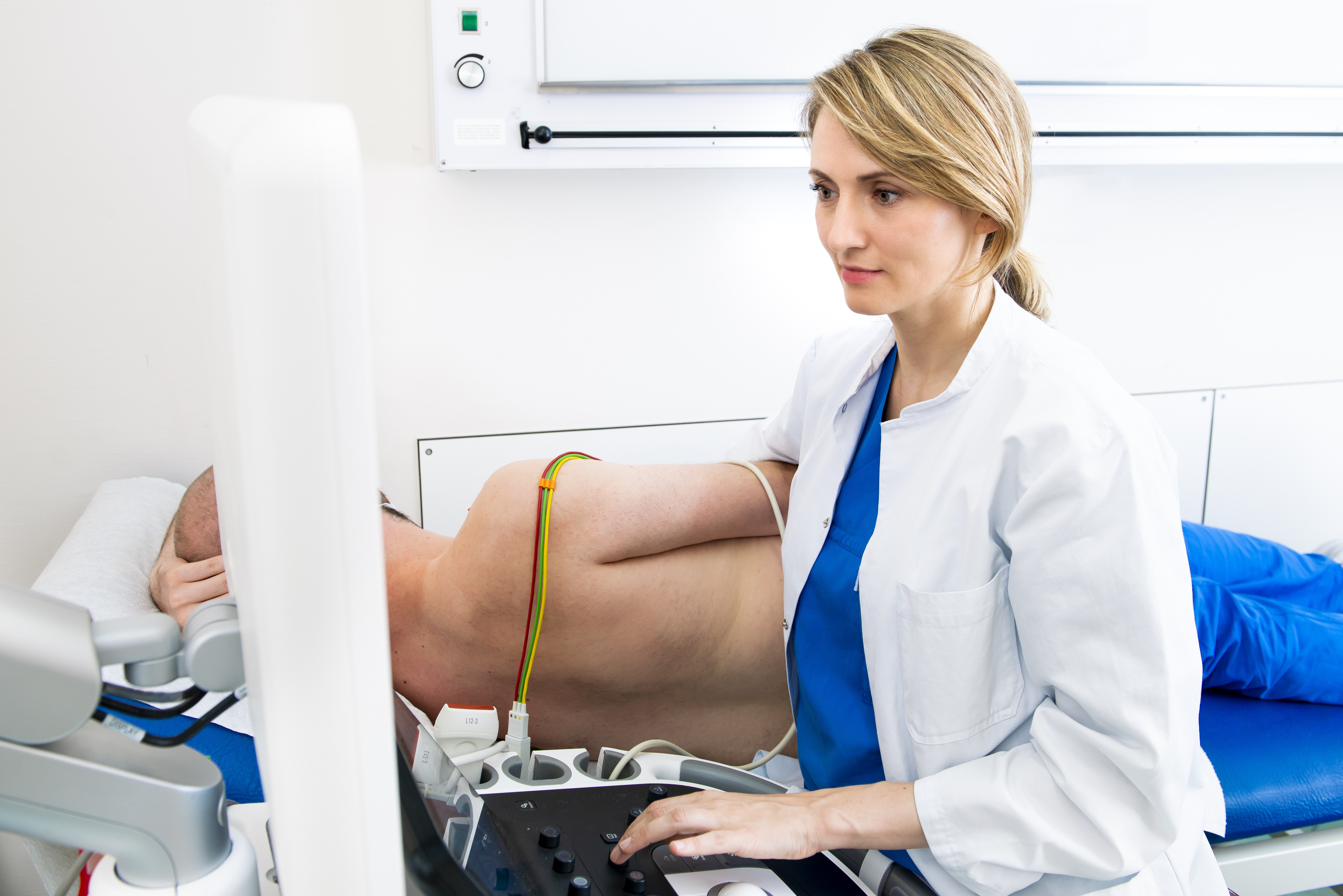
Stress echocardiography is an accompanying echocardiography during physical exertion of the patient. This can be generated on a bicycle ergometer or treadmill or simulated using medication.
Doctors use this method in particular to examine changes in wall movement under stress to determine whether a vascular repair (bypass or dilatation, possibly with a vascular support) is necessary. Stress echocardiography is also used to assess valve defects under stress.
Stress echocardiography is used to assess heart valve defects, among other things.
© DHZC
Stress echocardiography is used to assess heart valve defects, among other things.
© DHZC
Contrast echocardiography
Contrast echocardiography is characterised by the prior administration of a contrast agent: A well-tolerated liquid is introduced into the patient's vein and enters the heart with the blood. It stands out from the surrounding blood in the ultrasound image and enables the doctor to accurately assess the blood flow.
This enables the doctor to diagnose malformations and diseases of the heart, such as valve defects or narrowing of the coronary arteries.
Duplex sonography (vascular ultrasound)
Duplex sonography (vascular ultrasound) is used in the neck area. It is used to visualise the arteries supplying the brain (so-called ‘carotid Doppler’), the arteries of the arm and leg and the veins, e.g. in the case of deep vein thrombosis (‘venous Doppler’). Duplex sonography can also be carried out after interventions (‘peripheral Doppler’).
In addition to assessing the vessel, the blood flow velocity is also measured using the so-called Doppler effect. Carotid and brachial Doppler are performed preoperatively and in cases of vascular constriction. Peripheral Dopplers are carried out in cases of vasoconstriction, thrombosis, fistulas or vascular injuries.