Congenital tricuspid valve disorders
Congenital tricuspid valve disease affects the heart valve located between the right atrium and the right ventricle. This valve is called the tricuspid valve and ensures that blood can only flow forward through the heart and not backward. In tricuspid valve disease, the valve may either leak (“insufficient”) or be too narrow (“stenotic”). A combination of both conditions is also possible.
Tricuspid valve insufficiency (TI) and tricuspid valve stenosis (TS) can be congenital and occur without other heart defects, or they can develop in the course of a congenital or acquired heart disease. Isolated TS is very rare. It usually occurs in combination with TI.
A special form of tricuspid valve disease is the so-called Ebstein's anomaly.
Cause
Normally, oxygen-poor blood flows from the right atrium to the right ventricle via the tricuspid valve. From there, it is pumped into the pulmonary circulation, where it is enriched with oxygen. Both tricuspid valve insufficiency and tricuspid valve stenosis cause a backlog of oxygen-poor blood. The right atrium becomes stretched, which can promote the development of cardiac arrhythmias.
Risk factors
The cause of congenital tricuspid valve insufficiency is unknown. In the field of congenital heart defects, tricuspid valve insufficiency often occurs later in patients with various congenital heart defects, such as patients with Tetralogy of Fallot, truncus arteriosus and other malformations of the aortic arch and transposition of the great arteries. Patients who have undergone closure of a ventricular septal defect or an atrioventricular septal defect may also develop TI later on.
In the context of the aforementioned malformations, tricuspid valve insufficiency often occurs due to volume overload of the right ventricle. This leads to dilation of the tricuspid valve annulus and, consequently, to valve leakage. Acquired TI can be caused, for example, by infectious endocarditis and rheumatic heart disease, as well as being a “side effect” of a catheter procedure in the heart.
Isolated tricuspid valve stenosis is usually caused by rheumatic heart disease. In this case, several heart valves are often affected.
Symptoms
The symptoms of a child or adolescent with tricuspid valve disease vary depending on the severity of the TI or TS. An additional or previously corrected heart defect may also contribute to the symptoms.
Common symptoms of tricuspid valve insufficiency and tricuspid valve stenosis include general weakness and reduced cardiovascular capacity, an increase in the circumference of the abdomen and/or legs due to edema, prominent neck veins, and an irregular heartbeat. We recommend treatment of tricuspid valve disease to prevent long-term consequences such as irreversible damage to the right heart and chronic cardiac arrhythmias.
Tricuspid valve regurgitation is often treated by reconstructing the leaky heart valve. This procedure is usually performed surgically using a heart-lung machine.
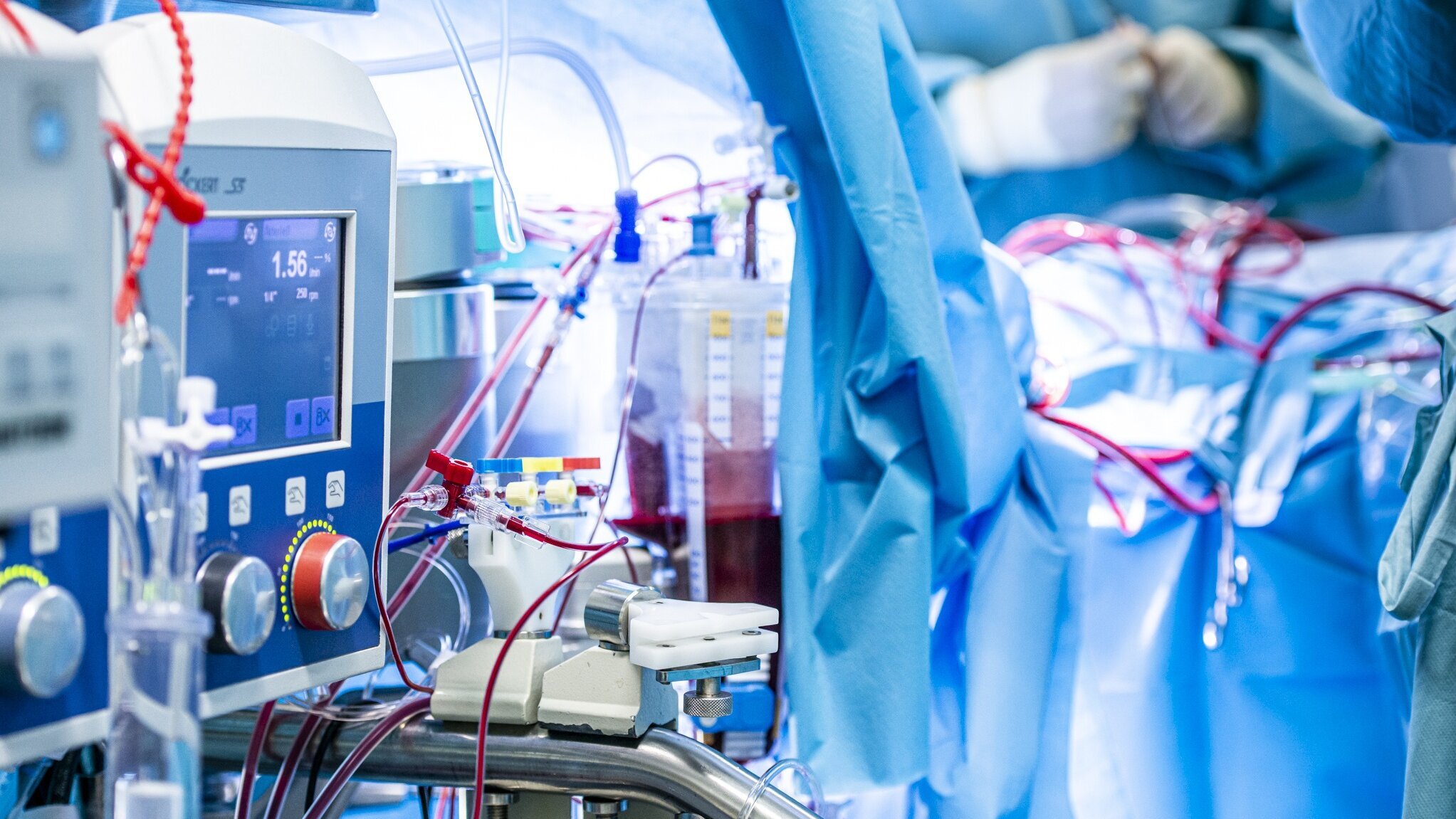
Tricuspid valve regurgitation is often treated by reconstructing the leaky heart valve. This procedure is usually performed surgically using a heart-lung machine.
Therapy
Tricuspid valve reconstruction for congenital malformations is usually corrected surgically using a heart-lung machine and must always be performed under general anesthesia.
There are medications that can support your child's heart until corrective surgery and alleviate the symptoms. However, these medications have no effect on heart valve function.
The incision that the pediatric heart surgeon must make during the operation runs vertically between the nipples. During the operation, the diseased tricuspid valve is reconstructed or, if this is not possible, replaced with a biological prosthesis. In tricuspid valve reconstruction, special rings or bands may be used, which are sutured around the enlarged tricuspid valve ring to reduce it to its normal size and thus enable the valve to close again. This preserves your child's valve. This option can be used for tricuspid valve insufficiency.
If tricuspid valve stenosis is present or the valve leaflets are so deformed that reconstruction does not appear promising, valve replacement is an option. There are two options available for tricuspid valve replacement using a prosthesis: biological prostheses made from porcine or bovine pericardium (i.e., heart sac) and mechanical prostheses made from plastic. Biological prostheses have proven effective in the tricuspid valve position because, unlike mechanical prostheses, they are less prone to blood clot formation. Another advantage of biological prostheses is that lifelong therapy with blood-thinning medication is not necessary if there are no cardiac arrhythmias.
Valve replacement surgery can also be performed interventionally using catheter technology. However, this procedure is used less frequently in children because children and their hearts are still growing and the prosthesis has no growth potential.
Therapy at the DHZC
Our clinic has more than 30 years of experience in reconstructing congenital tricuspid valve diseases and is a specialized center for the reconstruction of Ebstein's anomaly, among other conditions. Excellent results in the treatment of this malformation are only possible if, in addition to pure surgical expertise, extensive experience in anesthesia and intensive care is also available.
For example, it is very important that patients are weaned off artificial ventilation early after the procedure. For this purpose, there is a specially designed concept for these patients (ERAS: Enhanced recovery after surgery). This means that your child will be weaned off the ventilator while still in the operating room and will be able to breathe independently when they arrive at the pediatric intensive care unit. In the past, this has contributed to shorter stays in the intensive care unit and faster discharge home.
In some cases, minimally invasive access is also possible in adolescents and adults from the right side of the chest between the ribs. In order to operate using this approach, it is necessary to rule out the possibility of another defect in the heart that requires surgery. In some cases, but expressly not in all cases, this allows an incision in the middle of the chest and cutting through the sternum to be avoided.
Further information can be found in our externally validated quality assurance standards and annual quality reports.
Forecast
After successful tricuspid valve surgery, your child can usually resume normal physical activity. Immediately after the operation, it may be necessary to refrain from strenuous activity for a short period of time until healing is complete. Depending on the function of the right heart, some children may need to avoid strenuous activity in general. Your pediatric cardiologist will advise you individually on this.
Even after successful tricuspid valve surgery, you should take your child for regular follow-up appointments with your pediatric cardiologist or, later on, with a cardiologist specializing in adults with congenital heart defects (EMAH) in order to detect late effects such as atrial arrhythmias or renewed valve leakage in good time.
For about six months after tricuspid valve surgery, your child will need to take preventive antibiotics for certain procedures, such as dental work. This procedure is called “endocarditis prophylaxis” and is intended to prevent bacteria that enter the bloodstream during dental procedures from settling in the heart that has undergone surgery. Your pediatric cardiologist will inform you individually whether this endocarditis prophylaxis needs to be continued for more than six months.
The outlook in terms of life expectancy and quality of life depends on your child's right heart function and any additional heart defects. In some cases, even if the tricuspid valve reconstruction is initially successful, leakage may recur. This may require further surgery, which can have a negative impact on your child's quality of life. Cardiac arrhythmias may also develop later on.
Even if the tricuspid valve surgery is successful, leakage may recur over time or, after replacement with a prosthesis, prosthesis degeneration may occur. In both cases, further surgery may be necessary. Cardiac arrhythmias may also occur and may require treatment by catheter intervention.