Aortic surgery
The Deutsches Herzzentrum der Charité is one of the largest centres for aortic surgery in Germany in terms of case numbers.
More than 400 aortic surgeries are performed annually. After a thorough examination, specialised physicians decide on the optimal treatment method for each patient.
The often complex and complicated procedures and the follow-up care require experienced and well-rehearsed interdisciplinary teams, which are available at the DHZC around the clock. Doctors at the DHZC have contributed to the development of numerous novel surgical procedures in minimally invasive aortic surgery.
Surgery for aortic aneurysms
If an aortic aneurysm is diagnosed, normal blood pressure levels should be achieved first (with medication if necessary). If the aortic diameter is still below the recommended level for surgery, regular check-ups are also very important in addition to optimal blood pressure control. Minimising risk factors (e.g. quitting smoking) is also recommended, as this slows the growth of the aneurysm.
Ascending aorta (aorta ascendens)
The diameter of the aneurysm plays an important role in deciding on the form of treatment. In the case of an aneurysm of the ascending aorta, surgery is recommended if the diameter of the vessel is 55 millimetres or more, or if the aortic diameter is increasing rapidly (by more than 10 millimetres per year). If risk factorsare present, for example a familial predisposition, a rapid increase in size, aortic valve weakness or a desire to become pregnant, surgery should be performed starting at an aortic diameter of 45 millimetres.
For patients with connective tissue diseases of the aorta (e.g. Marfan syndrome), replacement of the ascending aorta is recommended from a diameter of 50 millimetres. In open-chest surgery (open surgery), a vascular prosthesis (sometimes with an aortic valve prosthesis) is inserted.
Descending aorta (aorta descendens)
For aneurysms of the descending aorta requiring treatment (diameter of 55 mm or more), a stent graft can be inserted using a minimally invasive technique (TEVAR; thoracic endovascular aortic repair). The prerequisite for this is that the aneurysm is a sufficient distance from the branching renal and pelvic vessels. Large aneurysms may not be suitable for such an intervention. In this case, open surgery must be performed. In the case of connective tissue disease, conventional surgical therapy is also necessary. Depending on the location of the aneurysm, a tubular or so-called Y-prosthesis is used at the level of the aneurysm.
Today, many aneurysms can be treated with endoprostheses without open surgery. In this minimally invasive procedure, known as endovascular implantation, the prosthesis is inserted through a small incision in the patient's groin or arm and advanced to the site of the aneurysm, where it is then deployed.
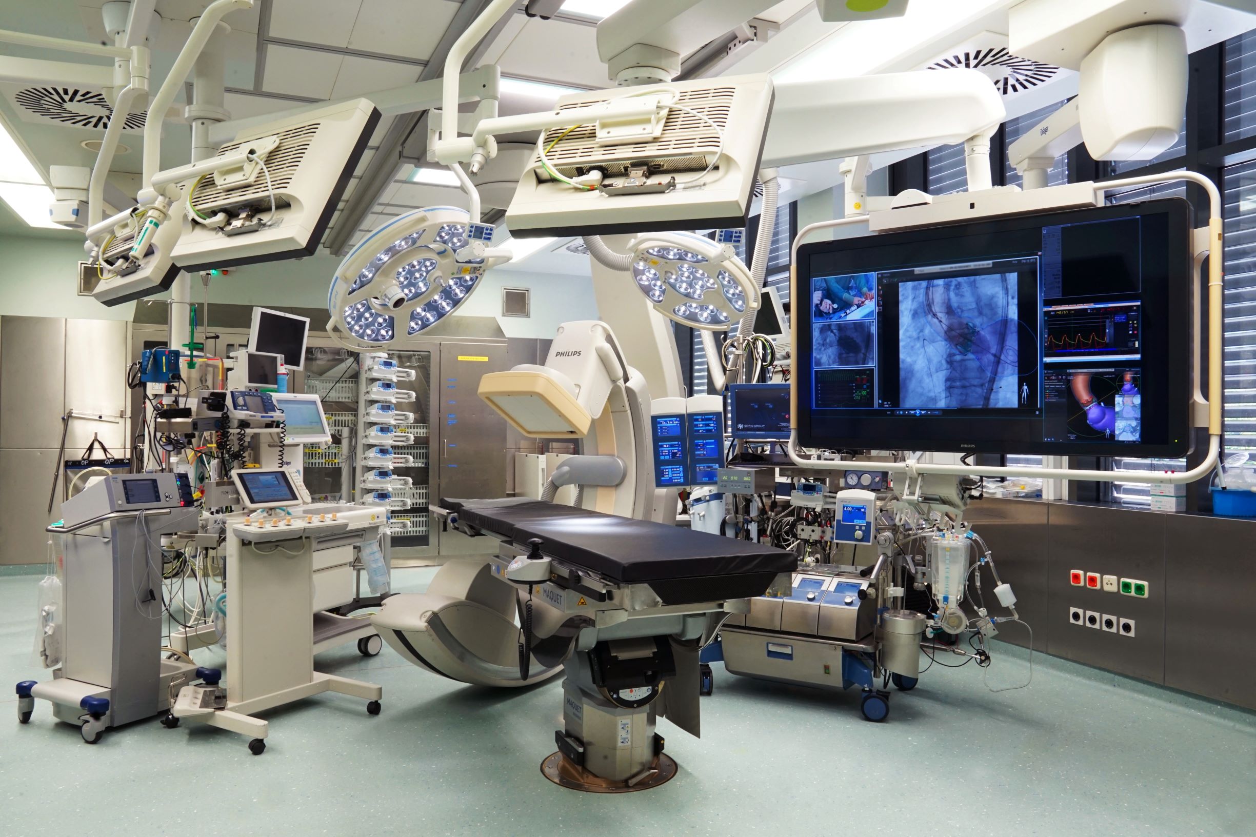
The latest surgical technology
More than 400 aortic operations are performed at the Deutsches Herzzentrum der Charité every year (Image: DHZC/Külker).
The latest surgical technology
More than 400 aortic operations are performed at the Deutsches Herzzentrum der Charité every year (Image: DHZC/Külker).
Aortic valve repair and replacement
In the case of a type A aortic dissection, the aortic valve and the coronary arteries may also be affected. The aim is always to preserve as many of the body's own structures as possible. Accordingly, reconstruction of the heart valves is preferred. If the valve function cannot be restored using the body's own structures, the valve must be replaced. In such cases, either a mechanical or a biological heart valve can be used.
Mechanical heart valves last a very long time, but the patient must take blood-thinning medication for the rest of his or her life. Biological valves are usually made from pericardium tissue taken from cows or from pig heart valves. With this type of prosthetic valve, the patient only needs to take blood-thinning medication during the first few weeks after surgery. However, these valves do not last forever and usually have to be replaced after about 10 to 15 years.
You can find out more about the treatment of aortic valve disease here.
During surgery on the ascending aorta or aortic arch, blood flow must be diverted during the operation. A heart-lung machine is used for this. The focus here is on two points:
Vital organs, primarily the brain, must continue to be supplied with oxygen-rich blood during the operation. The surgical area must be free of blood. To ensure this, the heart-lung machine takes over the patient's circulation and ensures an adequate supply of oxygen-rich blood.
During the operation, the heart stops beating under controlled conditions. A ‘stopped heart’ needs less oxygen to maintain the heart muscle cells. This gives the surgeon a window of time to treat the aortic disease. The heart-lung machine takes over the pumping function of the heart during this time.
A second possibility for reducing the oxygen requirement of the cells is hypothermia, or cooling the body temperature down to around 28 degrees Celsius. This is particularly beneficial for brain cells, in addition to heart muscle cells, which react to the reduced oxygen supply with equally reduced oxygen consumption.
To protect the brain cells even better from damage caused by a lack of oxygen (neuroprotection), there is the procedure of selective antegrade cerebral perfusion. In this procedure, vessels supplying the brain are additionally supplied with oxygen-rich blood from the heart-lung machine.
During surgery for aortic aneurysms and dissections, the diseased vessel is replaced by a prosthesis. In most cases, this prosthesis is made of plastic
(image: DHZC/Külker).
During surgery for aortic aneurysms and dissections, the diseased vessel is replaced by a prosthesis. In most cases, this prosthesis is made of plastic
(image: DHZC/Külker).
Surgical technique
In the case of aortic aneurysm or dissection surgery, the affected vessel is replaced with a prosthesis. This prosthesis is usually made of plastic. If the aorta or surrounding tissue is infected, homografts (removed with other organs during organ donation) or biological prostheses made of pericardium tissue may be used.
Conventional surgery
The way in which aortic surgery is performed depends on which segments of the aorta are affected. In the case of aneurysms in the aorta close to the heart and in the aortic arch, the sternum is cut and the patient's rib cage opened. The descending part of the aorta is accessed via an incision between the ribs. If the abdominal aorta is also affected, the incision is extended to the abdominal wall. If there is an aneurysm of the abdominal aorta below the renal arteries, this is accessed by opening the abdominal cavity.
The affected section of an aortic dissection or aneurysm is replaced by a vascular prosthesis. Depending on the location, this can be done using interventional (TEVAR) or open surgery. The prosthesis is an elastic tube made of synthetic fibres.
Rehabilitation
Rehabilitation after aortic surgery is typically divided into three phases:
- Early rehabilitation begins in the hospital and includes early mobilisation and physiotherapy.
- The subsequent curative treatment (AHB) normally takes place on an inpatient basis in the case of aortic and heart surgery, but can also be provided on an outpatient basis (as needed). Exercise therapy, physical therapy, psychosomatic care (e.g. to reduce anxiety) and health education (e.g. to manage risk factors) are all part of the programme.
- This is followed by re-integration (professional and non-professional). The aim here is to slowly increase the workload and to develop strategies for reducing and avoiding stress.
Regular check-ups
Patients with aortic disease require lifelong care, regardless of how the disease is treated (medically, interventionally or surgically). The frequency and extent of the check-ups depend on the type of procedure and aortic disease. The purpose of follow-up care during the first year is to monitor the success of the therapy and to quickly identify and eliminate any complications that may be associated with the procedure. Clinical examinations and imaging techniques (such as echocardiography, CT angiography, etc.) are required for follow-up care. Regular visits to the family doctor are also recommended.
Auto fahren
Innerhalb der ersten sechs Wochen nach der Operation sollten Sie auf das Fahren von Auto, Fahrrad und weiteren Fahrzeugen komplett verzichten. Da die Operation am Brustkorb stattfindet, ist nach dem Eingriff besonders darauf zu achten, das Brustbein so gut wie möglich zu schonen. Auch als Bei- oder Mitfahrer sollten Sie stets den Sicherheitsgurt anlegen.
Medikamente
Eine der wichtigsten Säulen in der Therapie von Aortenaneurysmen und Aortendissektionen ist neben der chirurgischen die medikamentöse Therapie. Risikofaktoren für das Herz-Kreislauf-System, wie Bluthochdruck oder zu hohe Blutzuckerwerte, sollten behoben und möglichst engmaschig kontrolliert werden. In Abhängigkeit von der Art der Aortenerkrankung und deren Therapie kann eine lebenslange Behandlung mit Thrombozytenaggregationshemmern (Blutplättchen-Hemmer) erforderlich sein. Hierfür eignet sich die Einnahme von ASS 100 mg einmal täglich. In besonderen Fällen kann auch eine Therapie mit Blutgerinnungshemmern, wie beispielsweise Marcumar® oder Falithrom®, notwendig sein.
Lebensführung
Da sowohl Aortenaneurysmen als auch -dissektionen als Folge von arteriosklerotischen Bindegewebsveränderungen entstehen können, gilt es nach dem Eingriff an der Aorta alle Risikofaktoren hierfür möglichst zu meiden oder zu reduzieren. Rauchen und Bluthochdruck sind hierbei die bedeutendsten Faktoren. Körperliche Aktivitäten und Sport können Sie etwa drei Monate nach der Operation beziehungsweise nach Abschluss der knöchernen Heilung des Brustbeins wiederaufnehmen.
Um Blutdruckspitzen zu vermeiden, sollte auf eine möglichst konstante Belastung geachtet werden. Aktivitäten mit gleichmäßiger Belastung sind unter anderem Schwimmen, Radfahren, Joggen und Walken. Krafttraining sollte nur in moderaten Maßen durchgeführt werden. Nach einem Aorteneingriff ist das Sexualleben nicht beeinträchtigt. Allerdings sollten auch hier übermäßige körperliche Anstrengungen vermieden werden, damit es zu keinen erhöhten Blutdruckwerten kommt.
Reisen und Wellness
Angesichts des Umfangs des Eingriffs und die anschließende Nachsorge sollten größere Reisen frühestens drei Monate nach der Operation unternommen werden. Versorgen Sie sich vor der Reise mit ausreichend Medikamenten und führen Sie Kopien wichtiger Unterlagen bei sich. Hierzu zählen Arztbrief, Prothesen-Ausweis usw. Achten Sie besonders in den ersten Monaten nach der Operation darauf, auf das Heben und Tragen schwerer Gegenstände (z.B. Reisegepäck) zu verzichten.
Auch hier gilt es, große körperliche Belastungen zu vermeiden. Höhenaufenthalte bis 2.000 Meter über dem Meeresspiegel stellen in der Regel kein Problem dar. Aufgrund der Wundheilung sollten mindestens drei Monate nach dem Eingriff abgewartet werden, bevor Sie eine Sauna oder ein Schwimmbad besuchen.
Berufsleben
Nach der erfolgreichen Therapie besteht in der Regel eine Arbeitsunfähigkeit für etwa drei Monate bzw. bis zum Abschluss der knöchernen Heilung. Im Verlauf wird, individuell und abhängig vom Genesungsgrad sowie ausgeübten Beruf, die Berufsfähigkeit eingestuft. Konnte eine Aortenerkrankung durch die Operation vollständig beseitigt werden, ist die Teilhabe am Leben in der Gemeinschaft meist unbeeinträchtigt möglich, sodass sich keine Behinderung aufgrund der Aortenoperation ergibt. Große, noch bestehende Aortenaneurysmen und chronische Aortendissektionen können allerdings zu Beeinträchtigungen im (Berufs-)Alltag führen und somit eine Behinderung darstellen.
Genetische Untersuchung
Aortenaneurysmen und -dissektionen können in Verbindung mit genetisch bedingten, angeborenen Bindegewebserkrankungen bzw. Syndromen vorkommen. Deren Ausprägung kann allerdings sehr unterschiedlich sein. Wann eine humangenetische Untersuchung sinnvoll ist, muss stets individuell entschieden werden. Empfohlen werden diese Untersuchungen bei relativ jungen Patient:innen mit einer Aortenerkrankung, denn hierbei können sich Konsequenzen für deren Nachsorge und auch für die nahen Verwandten ergeben.
Das Marfan-Zentrum an der Charité und dem DHZC ging aus den seit über zwanzig Jahren bestehenden speziellen Marfan-Sprechstunden der Humangenetik an der Charité und der Herzchirurgie am ehemaligen Deutschen Herzzentrum Berlin (DHZB) hervor. Eine gesetzliche Regelung (§ 116b SGB V) für die ambulante Betreuung von Patient:innen mit seltenen Erkrankungen wie dem Marfan-Syndrom ermöglicht es, die komplexe Betreuung und Behandlung unter einem Dach anzubieten. Hier bündelt sich die Erfahrung der Mediziner:innen. Idealerweise können die Patient:innen alle erforderlichen Termine mit geringem zeitlichem Aufwand am Marfan-Zentrum wahrnehmen.
About the author
Dr. med. Semih Buz works as a senior physician at the Department of Cardiac, Thoracic and Vascular Surgery at the German Heart Centre of the Charité (DHZC). He is head of the vascular surgery department.